 Movie
Movie Controller

[English] 日本語
 Yorodumi
- PDB-1ggp: CRYSTAL STRUCTURE OF TRICHOSANTHES KIRILOWII LECTIN-1 AND ITS REL... -
+ Open data

- Basic information
| Entry | Database: PDB / ID: 1ggp | ||||||
|---|---|---|---|---|---|---|---|
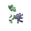
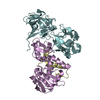
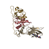
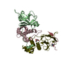
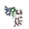
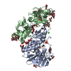
| Title | CRYSTAL STRUCTURE OF TRICHOSANTHES KIRILOWII LECTIN-1 AND ITS RELATION TO THE TYPE 2 RIBOSOME INACTIVATING PROTEINS | ||||||
 Components Components |
| ||||||
 Keywords Keywords | SUGAR BINDING PROTEIN / TRICHOSANTHES KIRILOWII / LECTIN | ||||||
| Function / homology |  Function and homology information Function and homology informationrRNA N-glycosylase / rRNA N-glycosylase activity / defense response / toxin activity / negative regulation of translation Similarity search - Function | ||||||
| Biological species |  Trichosanthes kirilowii (Chinese cucumber) Trichosanthes kirilowii (Chinese cucumber) | ||||||
| Method |  X-RAY DIFFRACTION / MOLECULAR REPLACEMENT / Resolution: 2.7 Å X-RAY DIFFRACTION / MOLECULAR REPLACEMENT / Resolution: 2.7 Å | ||||||
 Authors Authors | Li, M. / Chai, J.J. / Wang, Y.P. / Wang, K.Y. / Bi, R.C. | ||||||
 Citation Citation | Journal: PROTEIN PEPT.LETT. / Year: 2003 Title: Crystal Structure of Trichosanthes Kirilowii Lectin-1 and its Relation to the Type 2 Ribosome Inactivating Proteins Authors: Li, M. / Chai, J.J. / Wang, Y.P. / Wang, K.Y. / Bi, R.C. #1: Journal: Acta Crystallogr.,Sect.D / Year: 2000Title: Molecular-Replacement Studies of Trichosanthes Kirilowii Lectin-1: a Structure Belonging to the Family of Type 2 Ribosome-inactivating Proteins. Authors: Li, M. / Wang, Y.P. / Chai, J.J. / Wang, K.Y. / Bi, R.C. | ||||||
| History |
|
- Structure visualization
| Structure viewer | Molecule: MolmilJmol/JSmol |
|---|
- Downloads & links
-Download
| PDBx/mmCIF format | 1ggp.cif.gz | 92.9 KB | Display | PDBx/mmCIF format |
|---|---|---|---|---|
| PDB format | pdb1ggp.ent.gz | 70.9 KB | Display | PDB format |
| PDBx/mmJSON format | 1ggp.json.gz | Tree view | PDBx/mmJSON format | |
| Others |  Other downloads Other downloads |
-Validation report
| Arichive directory | https://data.pdbj.org/pub/pdb/validation_reports/gg/1ggpftp://data.pdbj.org/pub/pdb/validation_reports/gg/1ggp | HTTPS FTP |
|---|
-Related structure data
| Related structure data |  1abrS S: Starting model for refinement |
|---|---|
| Similar structure data |


-Links
 PDBj
PDBj

- Assembly
| Deposited unit | 
| ||||||||
|---|---|---|---|---|---|---|---|---|---|
| 1 |
| ||||||||
| Unit cell |
|
-Components
| #1: Protein | Mass: 24383.979 Da / Num. of mol.: 1 / Source method: isolated from a natural source Source: (natural) Trichosanthes kirilowii (Chinese cucumber)References: UniProt: Q7SIF0 |
|---|---|
| #2: Protein | Mass: 26645.523 Da / Num. of mol.: 1 / Source method: isolated from a natural source Source: (natural) Trichosanthes kirilowii (Chinese cucumber)References: UniProt: Q7SIF1 |
| #3: Water | ChemComp-HOH /  Mass: 18.015 Da / Num. of mol.: 12 / Source method: isolated from a natural source / Formula: H2O Mass: 18.015 Da / Num. of mol.: 12 / Source method: isolated from a natural source / Formula: H2O |
| Has protein modification | Y |
-Experimental details
-Experiment
| Experiment | Method: X-RAY DIFFRACTION / Number of used crystals: 1 |
|---|
- Sample preparation
| Crystal | Density Matthews: 2.78 Å3/Da / Density % sol: 55.82 % |
|---|---|
| Crystal grow | pH: 6 Details: Using the hanging-drop method, in which the droplets consisted of equal volume protein solution (4.2mg/ml) and the reservoir solution containing 15% PEG 8000, 0.5M Li2SO4 and 0.1M sodium ...Details: Using the hanging-drop method, in which the droplets consisted of equal volume protein solution (4.2mg/ml) and the reservoir solution containing 15% PEG 8000, 0.5M Li2SO4 and 0.1M sodium cacodylate buffer (pH 6.5). , pH 6.0 |
-Data collection
| Diffraction | Mean temperature: 293 K |
|---|---|
| Diffraction source | Source: ROTATING ANODE / Type: RIGAKU RU200 / Wavelength: 1.5418 |
| Detector | Type: MARRESEARCH / Detector: IMAGE PLATE |
| Radiation | Protocol: SINGLE WAVELENGTH / Monochromatic (M) / Laue (L): M / Scattering type: x-ray |
| Radiation wavelength | Wavelength: 1.5418 Å / Relative weight: 1 |
| Reflection | Resolution: 2.7→30 Å / Num. obs: 12480 / % possible obs: 74 % / Redundancy: 2.9 % / Biso Wilson estimate: 15.8 Å2 / Rsym value: 0.088 / Net I/σ(I): 7.4 |
| Reflection shell | Resolution: 2.7→2.8 Å / Redundancy: 2.95 % / Mean I/σ(I) obs: 2 / Rsym value: 0.37 / % possible all: 68 |
- Processing
| Software |
| ||||||||||||||||||||||||||||||||||||||||||||||||||||||||||||
|---|---|---|---|---|---|---|---|---|---|---|---|---|---|---|---|---|---|---|---|---|---|---|---|---|---|---|---|---|---|---|---|---|---|---|---|---|---|---|---|---|---|---|---|---|---|---|---|---|---|---|---|---|---|---|---|---|---|---|---|---|---|
| Refinement | Method to determine structure: MOLECULAR REPLACEMENT Starting model: 1ABR Resolution: 2.7→8 Å / Rfactor Rfree error: 0.012 / Data cutoff high absF: 10000000 / Data cutoff low absF: 0.001 / Isotropic thermal model: GROUP / Cross valid method: THROUGHOUT / σ(F): 2
| ||||||||||||||||||||||||||||||||||||||||||||||||||||||||||||
| Displacement parameters | Biso mean: 17.1 Å2
| ||||||||||||||||||||||||||||||||||||||||||||||||||||||||||||
| Refine analyze |
| ||||||||||||||||||||||||||||||||||||||||||||||||||||||||||||
| Refinement step | Cycle: LAST / Resolution: 2.7→8 Å
| ||||||||||||||||||||||||||||||||||||||||||||||||||||||||||||
| Refine LS restraints |
| ||||||||||||||||||||||||||||||||||||||||||||||||||||||||||||
| LS refinement shell | Resolution: 2.7→2.86 Å / Rfactor Rfree error: 0.039 / Total num. of bins used: 6
| ||||||||||||||||||||||||||||||||||||||||||||||||||||||||||||
| Xplor file |
|
