 Movie
Movie Controller

[English] 日本語
 Yorodumi
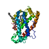
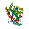
- PDB-1tfj: Crystal structure of Bovine Glycolipid transfer protein in comple... -
+ Open data

- Basic information
| Entry | Database: PDB / ID: 1tfj | ||||||
|---|---|---|---|---|---|---|---|
| Title | Crystal structure of Bovine Glycolipid transfer protein in complex with a fatty acid | ||||||
 Components Components | Glycolipid transfer protein | ||||||
 Keywords Keywords | LIPID TRANSPORT / Glycolipid | ||||||
| Function / homology |  Function and homology information Function and homology informationlipid transfer activity / ceramide 1-phosphate transfer activity / ceramide transport / ceramide 1-phosphate binding / glycolipid binding / intermembrane lipid transfer / lipid binding / cytosol Similarity search - Function | ||||||
| Biological species |  | ||||||
| Method |  X-RAY DIFFRACTION / SYNCHROTRON / MIR / Resolution: 1.61 Å X-RAY DIFFRACTION / SYNCHROTRON / MIR / Resolution: 1.61 Å | ||||||
 Authors Authors | Airenne, T.T. / Kidron, H. / West, G. / Nymalm, Y. / Mattjus, P. / Salminen, T.A. | ||||||
 Citation Citation | Journal: J.Mol.Biol. / Year: 2006 Title: Structural evidence for adaptive ligand binding of glycolipid transfer protein. Authors: Airenne, T.T. / Kidron, H. / Nymalm, Y. / Nylund, M. / West, G. / Mattjus, P. / Salminen, T.A. #1: Journal: Acta Crystallogr.,Sect.D / Year: 2004Title: Crystallization and X-ray analysis of bovine glycolipid transfer protein Authors: West, G. / Nymalm, Y. / Airenne, T.T. / Kidron, H. / Mattjus, P. / Salminen, T.T. | ||||||
| History |
|
- Structure visualization
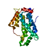
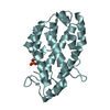
| Structure viewer | Molecule: MolmilJmol/JSmol |
|---|
- Downloads & links
-Download
| PDBx/mmCIF format | 1tfj.cif.gz | 59.5 KB | Display | PDBx/mmCIF format |
|---|---|---|---|---|
| PDB format | pdb1tfj.ent.gz | 43.4 KB | Display | PDB format |
| PDBx/mmJSON format | 1tfj.json.gz | Tree view | PDBx/mmJSON format | |
| Others |  Other downloads Other downloads |
-Validation report
| Summary document | 1tfj_validation.pdf.gz | 453.3 KB | Display | wwPDB validaton report |
|---|---|---|---|---|
| Full document | 1tfj_full_validation.pdf.gz | 455 KB | Display | |
| Data in XML | 1tfj_validation.xml.gz | 12.1 KB | Display | |
| Data in CIF | 1tfj_validation.cif.gz | 17.1 KB | Display | |
| Arichive directory | https://data.pdbj.org/pub/pdb/validation_reports/tf/1tfjftp://data.pdbj.org/pub/pdb/validation_reports/tf/1tfj | HTTPS FTP |
-Related structure data


-Links
 PDBj
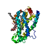
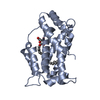
PDBj- Assembly
| Deposited unit | 
| ||||||||
|---|---|---|---|---|---|---|---|---|---|
| 1 |
| ||||||||
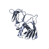
| Unit cell |
|
-Components
| #1: Protein | Mass: 25093.996 Da / Num. of mol.: 1 Source method: isolated from a genetically manipulated source Source: (gene. exp.)  |
|---|---|
| #2: Chemical | ChemComp-CL /   Mass: 35.453 Da / Num. of mol.: 1 / Source method: obtained synthetically / Formula: Cl Mass: 35.453 Da / Num. of mol.: 1 / Source method: obtained synthetically / Formula: Cl |
| #3: Chemical | ChemComp-DKA / 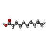  Mass: 172.265 Da / Num. of mol.: 1 / Source method: obtained synthetically / Formula: C10H20O2 Mass: 172.265 Da / Num. of mol.: 1 / Source method: obtained synthetically / Formula: C10H20O2 |
| #4: Chemical | ChemComp-GOL /   Mass: 92.094 Da / Num. of mol.: 1 / Source method: obtained synthetically / Formula: C3H8O3 Mass: 92.094 Da / Num. of mol.: 1 / Source method: obtained synthetically / Formula: C3H8O3 |
| #5: Water | ChemComp-HOH /  Mass: 18.015 Da / Num. of mol.: 151 / Source method: isolated from a natural source / Formula: H2O Mass: 18.015 Da / Num. of mol.: 151 / Source method: isolated from a natural source / Formula: H2O |
-Experimental details
-Experiment
| Experiment | Method: X-RAY DIFFRACTION / Number of used crystals: 1 |
|---|
- Sample preparation
| Crystal | Density Matthews: 2.1 Å3/Da / Density % sol: 41 % |
|---|---|
| Crystal grow | Temperature: 280 K / Method: vapor diffusion, hanging drop / pH: 6.6 Details: PEG 8000, MES, Sodium acetate, pH 6.6, VAPOR DIFFUSION, HANGING DROP, temperature 280K |
-Data collection
| Diffraction | Mean temperature: 100 K |
|---|---|
| Diffraction source | Source: SYNCHROTRON / Site: EMBL/DESY, HAMBURG  / Beamline: X13 / Wavelength: 0.804 Å / Beamline: X13 / Wavelength: 0.804 Å |
| Detector | Type: MARRESEARCH / Detector: CCD / Date: Oct 24, 2003 / Details: mirrors |
| Radiation | Monochromator: triangular / Protocol: SINGLE WAVELENGTH / Monochromatic (M) / Laue (L): M / Scattering type: x-ray |
| Radiation wavelength | Wavelength: 0.804 Å / Relative weight: 1 |
| Reflection | Resolution: 1.61→20 Å / Num. all: 26281 / Num. obs: 25509 / % possible obs: 97.1 % / Redundancy: 4.1 % / Rmerge(I) obs: 0.067 / Net I/σ(I): 12.16 |
| Reflection shell | Resolution: 1.61→1.7 Å / Redundancy: 3.8 % / Rmerge(I) obs: 0.414 / Mean I/σ(I) obs: 3.27 / Num. unique all: 3717 / % possible all: 95.1 |
- Processing
| Software |
| |||||||||||||||||||||||||||||||||||||||||||||||||||||||||||||||||||||||||||||||||||||||||||||||||||||||||||||||||||||||||||||
|---|---|---|---|---|---|---|---|---|---|---|---|---|---|---|---|---|---|---|---|---|---|---|---|---|---|---|---|---|---|---|---|---|---|---|---|---|---|---|---|---|---|---|---|---|---|---|---|---|---|---|---|---|---|---|---|---|---|---|---|---|---|---|---|---|---|---|---|---|---|---|---|---|---|---|---|---|---|---|---|---|---|---|---|---|---|---|---|---|---|---|---|---|---|---|---|---|---|---|---|---|---|---|---|---|---|---|---|---|---|---|---|---|---|---|---|---|---|---|---|---|---|---|---|---|---|---|
| Refinement | Method to determine structure: MIR / Resolution: 1.61→18.63 Å / Cor.coef. Fo:Fc: 0.963 / Cor.coef. Fo:Fc free: 0.942 / SU B: 1.955 / SU ML: 0.069 / Cross valid method: THROUGHOUT / ESU R: 0.1 / ESU R Free: 0.102 / Stereochemistry target values: MAXIMUM LIKELIHOOD / Details: HYDROGENS HAVE BEEN ADDED IN THE RIDING POSITIONS
| |||||||||||||||||||||||||||||||||||||||||||||||||||||||||||||||||||||||||||||||||||||||||||||||||||||||||||||||||||||||||||||
| Solvent computation | Ion probe radii: 0.8 Å / Shrinkage radii: 0.8 Å / VDW probe radii: 1.2 Å / Solvent model: BABINET MODEL WITH MASK | |||||||||||||||||||||||||||||||||||||||||||||||||||||||||||||||||||||||||||||||||||||||||||||||||||||||||||||||||||||||||||||
| Displacement parameters | Biso mean: 24.122 Å2
| |||||||||||||||||||||||||||||||||||||||||||||||||||||||||||||||||||||||||||||||||||||||||||||||||||||||||||||||||||||||||||||
| Refinement step | Cycle: LAST / Resolution: 1.61→18.63 Å
| |||||||||||||||||||||||||||||||||||||||||||||||||||||||||||||||||||||||||||||||||||||||||||||||||||||||||||||||||||||||||||||
| Refine LS restraints |
| |||||||||||||||||||||||||||||||||||||||||||||||||||||||||||||||||||||||||||||||||||||||||||||||||||||||||||||||||||||||||||||
| LS refinement shell | Resolution: 1.61→1.7 Å / Total num. of bins used: 20
|
