 Movie
Movie Controller


+ Open data

- Basic information
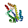
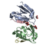
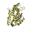
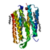
| Entry | Database: PDB / ID: 2bv7 | ||||||
|---|---|---|---|---|---|---|---|
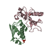
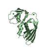
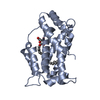
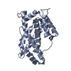
| Title | Crystal structure of GLTP with bound GM3 | ||||||
 Components Components | GLYCOLIPID TRANSFER PROTEIN | ||||||
 Keywords Keywords | LIPID TRANSPORT / GLYCOLIPID / LIGAND BINDING / ACETYLATION / TRANSPORT / GLYCOLIPID TRANSFER PROTEIN | ||||||
| Function / homology |  Function and homology information Function and homology informationglycolipid transfer activity / ceramide 1-phosphate transfer activity / ceramide transport / ceramide 1-phosphate binding / glycolipid binding / lipid transfer activity / intermembrane lipid transfer / lipid binding / cytosol Similarity search - Function | ||||||
| Biological species |  | ||||||
| Method |  X-RAY DIFFRACTION / SYNCHROTRON / MOLECULAR REPLACEMENT / Resolution: 1.79 Å X-RAY DIFFRACTION / SYNCHROTRON / MOLECULAR REPLACEMENT / Resolution: 1.79 Å | ||||||
 Authors Authors | Kidron, H. / Airenne, T.T. / Nymalm, Y. / Nylund, M. / West, G. / Mattjus, P. / Salminen, T.A. | ||||||
 Citation Citation | Journal: J.Mol.Biol. / Year: 2006 Title: Structural Evidence for Adaptive Ligand Binding of Glycolipid Transfer Protein. Authors: Airenne, T.T. / Kidron, H. / Nymalm, Y. / Nylund, M. / West, G. / Mattjus, P. / Salminen, T.A. #1: Journal: Acta Crystallogr.,Sect.D / Year: 2004 Title: Crystallization and X-Ray Analysis of Bovine Glycolipid Transfer Protein Authors: West, G. / Nymalm, Y. / Airenne, T.T. / Kidron, H. / Mattjus, P. / Salminen, T.A. | ||||||
| History |
|
- Structure visualization
| Structure viewer | Molecule: MolmilJmol/JSmol |
|---|
- Downloads & links
-Download
| PDBx/mmCIF format | 2bv7.cif.gz | 59.7 KB | Display | PDBx/mmCIF format |
|---|---|---|---|---|
| PDB format | pdb2bv7.ent.gz | 41.7 KB | Display | PDB format |
| PDBx/mmJSON format | 2bv7.json.gz | Tree view | PDBx/mmJSON format | |
| Others |  Other downloads Other downloads |
-Validation report
| Arichive directory | https://data.pdbj.org/pub/pdb/validation_reports/bv/2bv7ftp://data.pdbj.org/pub/pdb/validation_reports/bv/2bv7 | HTTPS FTP |
|---|
-Related structure data
| Related structure data |  1tfjSC  1wbeC S: Starting model for refinement C: citing same article ( |
|---|---|
| Similar structure data |


-Links
 PDBj
PDBj- Assembly
| Deposited unit | 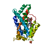
| ||||||||
|---|---|---|---|---|---|---|---|---|---|
| 1 |
| ||||||||
| Unit cell |
|
-Components
| #1: Protein | Mass: 23819.660 Da / Num. of mol.: 1 Source method: isolated from a genetically manipulated source Source: (gene. exp.)  | ||||||
|---|---|---|---|---|---|---|---|
| #2: Chemical |   Mass: 96.063 Da / Num. of mol.: 2 / Source method: obtained synthetically / Formula: SO4 Mass: 96.063 Da / Num. of mol.: 2 / Source method: obtained synthetically / Formula: SO4#3: Chemical | ChemComp-GM3 / |   Mass: 842.323 Da / Num. of mol.: 1 / Source method: obtained synthetically / Formula: C50H99NO8 Mass: 842.323 Da / Num. of mol.: 1 / Source method: obtained synthetically / Formula: C50H99NO8#4: Water | ChemComp-HOH / |  Mass: 18.015 Da / Num. of mol.: 119 / Source method: isolated from a natural source / Formula: H2O Mass: 18.015 Da / Num. of mol.: 119 / Source method: isolated from a natural source / Formula: H2OCompound details | ACCELERATE | |
-Experimental details
-Experiment
| Experiment | Method: X-RAY DIFFRACTION / Number of used crystals: 1 |
|---|
- Sample preparation
| Crystal | Density Matthews: 2.1 Å3/Da / Density % sol: 40.6 % |
|---|
-Data collection
| Diffraction | Mean temperature: 100 K |
|---|---|
| Diffraction source | Source: SYNCHROTRON / Site: ESRF  / Beamline: ID14-1 / Wavelength: 0.934 / Beamline: ID14-1 / Wavelength: 0.934 |
| Detector | Type: ADSC CCD / Detector: CCD / Date: Nov 8, 2004 |
| Radiation | Protocol: SINGLE WAVELENGTH / Monochromatic (M) / Laue (L): M / Scattering type: x-ray |
| Radiation wavelength | Wavelength: 0.934 Å / Relative weight: 1 |
| Reflection | Resolution: 1.8→25 Å / Num. obs: 115280 / % possible obs: 99.5 % / Observed criterion σ(I): -3 / Redundancy: 6 % / Rmerge(I) obs: 0.08 / Net I/σ(I): 16.6 |
| Reflection shell | Resolution: 1.8→1.9 Å / Redundancy: 6.14 % / Rmerge(I) obs: 0.52 / Mean I/σ(I) obs: 4.2 / % possible all: 99.7 |
- Processing
| Software |
| ||||||||||||||||||||||||||||||||||||||||||||||||||||||||||||||||||||||||||||||||||||||||||||||||||||||||||||||||||||||||||||||||||||||||||||||||||||||||||||||||||||||||||||||||||||||
|---|---|---|---|---|---|---|---|---|---|---|---|---|---|---|---|---|---|---|---|---|---|---|---|---|---|---|---|---|---|---|---|---|---|---|---|---|---|---|---|---|---|---|---|---|---|---|---|---|---|---|---|---|---|---|---|---|---|---|---|---|---|---|---|---|---|---|---|---|---|---|---|---|---|---|---|---|---|---|---|---|---|---|---|---|---|---|---|---|---|---|---|---|---|---|---|---|---|---|---|---|---|---|---|---|---|---|---|---|---|---|---|---|---|---|---|---|---|---|---|---|---|---|---|---|---|---|---|---|---|---|---|---|---|---|---|---|---|---|---|---|---|---|---|---|---|---|---|---|---|---|---|---|---|---|---|---|---|---|---|---|---|---|---|---|---|---|---|---|---|---|---|---|---|---|---|---|---|---|---|---|---|---|---|
| Refinement | Method to determine structure: MOLECULAR REPLACEMENT Starting model: PDB ENTRY 1TFJ Resolution: 1.79→52.2 Å / Cor.coef. Fo:Fc: 0.95 / Cor.coef. Fo:Fc free: 0.908 / SU B: 2.785 / SU ML: 0.09 / Cross valid method: THROUGHOUT / ESU R: 0.142 / ESU R Free: 0.149 / Stereochemistry target values: MAXIMUM LIKELIHOOD / Details: HYDROGENS HAVE BEEN ADDED IN THE RIDING POSITIONS.
| ||||||||||||||||||||||||||||||||||||||||||||||||||||||||||||||||||||||||||||||||||||||||||||||||||||||||||||||||||||||||||||||||||||||||||||||||||||||||||||||||||||||||||||||||||||||
| Solvent computation | Ion probe radii: 0.8 Å / Shrinkage radii: 0.8 Å / VDW probe radii: 1.2 Å / Solvent model: MASK | ||||||||||||||||||||||||||||||||||||||||||||||||||||||||||||||||||||||||||||||||||||||||||||||||||||||||||||||||||||||||||||||||||||||||||||||||||||||||||||||||||||||||||||||||||||||
| Displacement parameters | Biso mean: 22.82 Å2
| ||||||||||||||||||||||||||||||||||||||||||||||||||||||||||||||||||||||||||||||||||||||||||||||||||||||||||||||||||||||||||||||||||||||||||||||||||||||||||||||||||||||||||||||||||||||
| Refinement step | Cycle: LAST / Resolution: 1.79→52.2 Å
| ||||||||||||||||||||||||||||||||||||||||||||||||||||||||||||||||||||||||||||||||||||||||||||||||||||||||||||||||||||||||||||||||||||||||||||||||||||||||||||||||||||||||||||||||||||||
| Refine LS restraints |
|