Trafficking and processing of endosomal TLR / Scavenging by Class A Receptors / Regulation of Insulin-like Growth Factor (IGF) transport and uptake by Insulin-like Growth Factor Binding Proteins (IGFBPs) / Interleukin-4 and Interleukin-13 signaling / Post-translational protein phosphorylation / sarcoplasmic reticulum lumen / ERAD pathway / ATP-dependent protein folding chaperone / Hydrolases; Acting on acid anhydrides; Acting on acid anhydrides to facilitate cellular and subcellular movement / : ...Trafficking and processing of endosomal TLR / Scavenging by Class A Receptors / Regulation of Insulin-like Growth Factor (IGF) transport and uptake by Insulin-like Growth Factor Binding Proteins (IGFBPs) / Interleukin-4 and Interleukin-13 signaling / Post-translational protein phosphorylation / sarcoplasmic reticulum lumen / ERAD pathway / ATP-dependent protein folding chaperone / Hydrolases; Acting on acid anhydrides; Acting on acid anhydrides to facilitate cellular and subcellular movement / : / melanosome / protein folding / perinuclear region of cytoplasm / endoplasmic reticulum / ATP hydrolysis activity / ATP binding Similarity search - Function
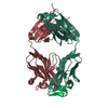
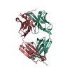
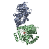
Endoplasmic reticulum targeting sequence. / Heat shock protein Hsp90, conserved site / Heat shock hsp90 proteins family signature. / Histidine kinase-like ATPase, C-terminal domain / Heat Shock Protein 90 / HSP90, C-terminal domain / Heat shock protein Hsp90, N-terminal / Heat shock protein Hsp90 family / Hsp90 protein / Histidine kinase-, DNA gyrase B-, and HSP90-like ATPase ...Endoplasmic reticulum targeting sequence. / Heat shock protein Hsp90, conserved site / Heat shock hsp90 proteins family signature. / Histidine kinase-like ATPase, C-terminal domain / Heat Shock Protein 90 / HSP90, C-terminal domain / Heat shock protein Hsp90, N-terminal / Heat shock protein Hsp90 family / Hsp90 protein / Histidine kinase-, DNA gyrase B-, and HSP90-like ATPase / Histidine kinase-, DNA gyrase B-, and HSP90-like ATPase / Histidine kinase-like ATPases / Histidine kinase/HSP90-like ATPase / Histidine kinase/HSP90-like ATPase superfamily / Ribosomal protein S5 domain 2-type fold / 2-Layer Sandwich / Alpha Beta Similarity search - Domain/homology
HETEROGEN ONLY FRAGMENTS OF PEG400 (PG4) WERE IDENTIFIED AND IN MANY CASES SEVERAL ATOMS WERE ...HETEROGEN ONLY FRAGMENTS OF PEG400 (PG4) WERE IDENTIFIED AND IN MANY CASES SEVERAL ATOMS WERE MISSING DUE TO LACK OF ELECTRON DENSITY. THE GAMMA PHOSPHATE IS DISORDERED IN THE ATP LIGAND.
Remark 999
SEQUENCE SEQUENCE DATABASE RESIDUES 287-327 WERE DELETED AND REPLACED BY 4 GLYCINES.
In the structure databanks used in Yorodumi, some data are registered as the other names, "COVID-19 virus" and "2019-nCoV". Here are the details of the virus and the list of structure data.
Jan 31, 2019. EMDB accession codes are about to change! (news from PDBe EMDB page)
EMDB accession codes are about to change! (news from PDBe EMDB page)
The allocation of 4 digits for EMDB accession codes will soon come to an end. Whilst these codes will remain in use, new EMDB accession codes will include an additional digit and will expand incrementally as the available range of codes is exhausted. The current 4-digit format prefixed with “EMD-” (i.e. EMD-XXXX) will advance to a 5-digit format (i.e. EMD-XXXXX), and so on. It is currently estimated that the 4-digit codes will be depleted around Spring 2019, at which point the 5-digit format will come into force.
The EM Navigator/Yorodumi systems omit the EMD- prefix.
Related info.:Q: What is EMD? / ID/Accession-code notation in Yorodumi/EM Navigator
Yorodumi is a browser for structure data from EMDB, PDB, SASBDB, etc.
This page is also the successor to EM Navigator detail page, and also detail information page/front-end page for Omokage search.
The word "yorodu" (or yorozu) is an old Japanese word meaning "ten thousand". "mi" (miru) is to see.
Related info.:EMDB / PDB / SASBDB / Comparison of 3 databanks / Yorodumi Search / Aug 31, 2016. New EM Navigator & Yorodumi / Yorodumi Papers / Jmol/JSmol / Function and homology information / Changes in new EM Navigator and Yorodumi
 Movie
Movie Controller

 Yorodumi
 Open data
 Basic information
 Components
 Keywords
 Function and homology information
 X-RAY DIFFRACTION /
  Authors
 Citation
 Structure visualization
 Downloads & links
 Other downloads


 PDBj
PDBj


 Assembly
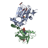


 Mass: 24.305 Da / Num. of mol.: 2 / Source method: obtained synthetically / Formula: Mg
 Mass: 507.181 Da / Num. of mol.: 2 / Source method: obtained synthetically / Formula: C10H16N5O13P3 / Comment: ATP, energy-carrying molecule*YM
 Mass: 194.226 Da / Num. of mol.: 5 / Source method: obtained synthetically / Formula: C8H18O5 / Comment: precipitant*YM
 Mass: 18.015 Da / Num. of mol.: 203 / Source method: isolated from a natural source / Formula: H2O
 Sample preparation
 Processing