 Movie
Movie Controller


+ Open data

- Basic information
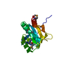
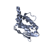
| Entry | Database: PDB / ID: 1qpv | ||||||
|---|---|---|---|---|---|---|---|
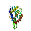
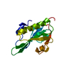
| Title | YEAST COFILIN | ||||||
 Components Components | YEAST COFILIN | ||||||
 Keywords Keywords | ACTIN-BINDING PROTEIN / THREE LAYERS:ALPHA/MIXED BETA/ALPHA / COFILIN FOLD | ||||||
| Function / homology |  Function and homology information Function and homology informationactin cortical patch / actin filament severing / Golgi to plasma membrane protein transport / actin filament depolymerization / actin filament organization / nuclear matrix / endocytosis / actin filament binding / actin cytoskeleton / plasma membrane / cytoplasm Similarity search - Function | ||||||
| Biological species |  | ||||||
| Method |  X-RAY DIFFRACTION / MOLECULAR REPLACEMENT / Resolution: 3 Å X-RAY DIFFRACTION / MOLECULAR REPLACEMENT / Resolution: 3 Å | ||||||
 Authors Authors | Fedorov, A.A. / Lappalainen, P. / Fedorov, E.V. / Drubin, D.G. / Almo, S.C. | ||||||
 Citation Citation | Journal: Nat.Struct.Biol. / Year: 1997 Title: Structure determination of yeast cofilin. Authors: Fedorov, A.A. / Lappalainen, P. / Fedorov, E.V. / Drubin, D.G. / Almo, S.C. #1: Journal: Embo J. / Year: 1997Title: Essential Functions and Actin-Binding Surfaces of Yeast Cofilin Revealed by Systematic Mutagenesis Authors: Lappalainen, P. / Fedorov, E.V. / Fedorov, A.A. / Almo, S.C. / Drubin, D.G. #2: Journal: Nature / Year: 1997Title: Cofilin Promotes Rapid Actin Filament Turnover in Vivo Authors: Lappalainen, P. / Drubin, D.G. | ||||||
| History |
|
- Structure visualization
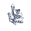
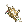
| Structure viewer | Molecule: MolmilJmol/JSmol |
|---|
- Downloads & links
-Download
| PDBx/mmCIF format | 1qpv.cif.gz | 36.8 KB | Display | PDBx/mmCIF format |
|---|---|---|---|---|
| PDB format | pdb1qpv.ent.gz | 25.3 KB | Display | PDB format |
| PDBx/mmJSON format | 1qpv.json.gz | Tree view | PDBx/mmJSON format | |
| Others |  Other downloads Other downloads |
-Validation report
| Arichive directory | https://data.pdbj.org/pub/pdb/validation_reports/qp/1qpvftp://data.pdbj.org/pub/pdb/validation_reports/qp/1qpv | HTTPS FTP |
|---|
-Related structure data
| Related structure data |  1cfyC  1cofSC C: citing same article ( S: Starting model for refinement |
|---|---|
| Similar structure data |


-Links
 PDBj
PDBj
- Assembly
| Deposited unit | 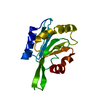
| ||||||||
|---|---|---|---|---|---|---|---|---|---|
| 1 |
| ||||||||
| Unit cell |
| ||||||||
| Details | biological unit is monomer |
-Components
| #1: Protein | Mass: 15919.742 Da / Num. of mol.: 1 Source method: isolated from a genetically manipulated source Source: (gene. exp.) Production host:  |
|---|
-Experimental details
-Experiment
| Experiment | Method: X-RAY DIFFRACTION / Number of used crystals: 1 |
|---|
- Sample preparation
| Crystal | Density Matthews: 1.99 Å3/Da / Density % sol: 38 % |
|---|---|
| Crystal grow | Temperature: 290 K / Method: vapor diffusion, hanging drop / pH: 7.4 Details: PEG 4000 , pH 7.4, VAPOR DIFFUSION, HANGING DROP, temperature 290K |
-Data collection
| Diffraction | Mean temperature: 290 K |
|---|---|
| Diffraction source | Source: ROTATING ANODE / Type: RIGAKU RU200 / Wavelength: 1.5418 |
| Detector | Type: SIEMENS / Detector: AREA DETECTOR / Date: Feb 15, 1995 |
| Radiation | Monochromator: GRAPHITE CRYSTAL / Protocol: SINGLE WAVELENGTH / Monochromatic (M) / Laue (L): M / Scattering type: x-ray |
| Radiation wavelength | Wavelength: 1.5418 Å / Relative weight: 1 |
| Reflection | Resolution: 3→25.2 Å / Num. all: 2529 / Num. obs: 2377 / % possible obs: 89.1 % / Observed criterion σ(F): 0 / Observed criterion σ(I): 1 / Redundancy: 2.9 % / Biso Wilson estimate: 16.2 Å2 / Rmerge(I) obs: 0.083 / Net I/σ(I): 19.8 |
| Reflection shell | Resolution: 3→3.1 Å / Redundancy: 2.2 % / Rmerge(I) obs: 0.185 / Mean I/σ(I) obs: 4.9 / % possible all: 77.4 |
- Processing
| Software |
| |||||||||||||||||||||||||
|---|---|---|---|---|---|---|---|---|---|---|---|---|---|---|---|---|---|---|---|---|---|---|---|---|---|---|
| Refinement | Method to determine structure: MOLECULAR REPLACEMENT Starting model: 1COF Resolution: 3→8 Å / Isotropic thermal model: RESTRAINED / Cross valid method: THROUGHOUT / σ(F): 2 / σ(I): 0 / Stereochemistry target values: Engh & Huber
| |||||||||||||||||||||||||
| Refinement step | Cycle: LAST / Resolution: 3→8 Å
| |||||||||||||||||||||||||
| Refine LS restraints |
| |||||||||||||||||||||||||
| Xplor file | Serial no: 1 / Param file: PROTEIN_REP.PARAM / Topol file: TOPHCSDX.PRO |
