 Movie
Movie Controller

[English] 日本語
 Yorodumi
- PDB-2vik: REFINED STRUCTURE OF THE ACTIN-SEVERING DOMAIN VILLIN 14T, DETERM... -
+ Open data

- Basic information
| Entry | Database: PDB / ID: 2vik | ||||||
|---|---|---|---|---|---|---|---|
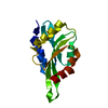
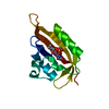
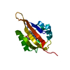
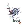
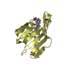
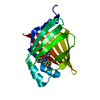
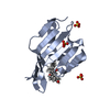
| Title | REFINED STRUCTURE OF THE ACTIN-SEVERING DOMAIN VILLIN 14T, DETERMINED BY SOLUTION NMR, MINIMIZED AVERAGE STRUCTURE | ||||||
 Components Components | VILLIN 14T | ||||||
 Keywords Keywords | ACTIN-BINDING PROTEIN / CAPPING PROTEIN / CALCIUM-BINDING PROTEIN / CYTOSKELETAL PROTEIN | ||||||
| Function / homology |  Function and homology information Function and homology informationregulation of actin nucleation / lysophosphatidic acid binding / regulation of lamellipodium morphogenesis / cytoplasmic actin-based contraction involved in cell motility / filopodium tip / positive regulation of actin filament bundle assembly / regulation of wound healing / actin filament severing / barbed-end actin filament capping / cysteine-type endopeptidase inhibitor activity involved in apoptotic process ...regulation of actin nucleation / lysophosphatidic acid binding / regulation of lamellipodium morphogenesis / cytoplasmic actin-based contraction involved in cell motility / filopodium tip / positive regulation of actin filament bundle assembly / regulation of wound healing / actin filament severing / barbed-end actin filament capping / cysteine-type endopeptidase inhibitor activity involved in apoptotic process / actin polymerization or depolymerization / actin filament depolymerization / actin filament capping / cellular response to hepatocyte growth factor stimulus / actin filament bundle / microvillus / positive regulation of epithelial cell migration / ruffle / actin filament polymerization / phosphatidylinositol-4,5-bisphosphate binding / cellular response to epidermal growth factor stimulus / response to bacterium / filopodium / epidermal growth factor receptor signaling pathway / actin filament binding / regulation of cell shape / lamellipodium / actin cytoskeleton / positive regulation of cell migration / calcium ion binding / protein homodimerization activity / cytoplasm Similarity search - Function | ||||||
| Biological species |  | ||||||
| Method | SOLUTION NMR / distance geometry | ||||||
 Authors Authors | Markus, M.A. / Matsudaira, P. / Wagner, G. | ||||||
 Citation Citation | Journal: Protein Sci. / Year: 1997 Title: Refined structure of villin 14T and a detailed comparison with other actin-severing domains. Authors: Markus, M.A. / Matsudaira, P. / Wagner, G. #1: Journal: Biochemistry / Year: 1996Title: Local Mobility within Villin 14T Probed Via Heteronuclear Relaxation Measurements and a Reduced Spectral Density Mapping Authors: Markus, M.A. / Dayie, K.T. / Matsudaira, P. / Wagner, G. #2: Journal: Protein Sci. / Year: 1994Title: Solution Structure of Villin 14T, a Domain Conserved Among Actin-Severing Proteins Authors: Markus, M.A. / Nakayama, T. / Matsudaira, P. / Wagner, G. #3: Journal: J.Biomol.NMR / Year: 1994Title: 1H, 15N, 13C and 13Co Resonance Assignments and Secondary Structure of Villin 14T, a Domain Conserved Among Actin-Severing Proteins Authors: Markus, M.A. / Nakayama, T. / Matsudaira, P. / Wagner, G. | ||||||
| History |
|
- Structure visualization
| Structure viewer | Molecule: MolmilJmol/JSmol |
|---|
- Downloads & links
-Download
| PDBx/mmCIF format | 2vik.cif.gz | 56.1 KB | Display | PDBx/mmCIF format |
|---|---|---|---|---|
| PDB format | pdb2vik.ent.gz | 40.6 KB | Display | PDB format |
| PDBx/mmJSON format | 2vik.json.gz | Tree view | PDBx/mmJSON format | |
| Others |  Other downloads Other downloads |
-Validation report
| Arichive directory | https://data.pdbj.org/pub/pdb/validation_reports/vi/2vikftp://data.pdbj.org/pub/pdb/validation_reports/vi/2vik | HTTPS FTP |
|---|
-Related structure data


-Links
 PDBj
PDBj
- Assembly
| Deposited unit | 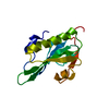
| |||||||||
|---|---|---|---|---|---|---|---|---|---|---|
| 1 |
| |||||||||
| NMR ensembles |
|
-Components
| #1: Protein | Mass: 14174.928 Da / Num. of mol.: 1 / Fragment: RESIDUES 1 - 126 Source method: isolated from a genetically manipulated source Source: (gene. exp.)  |
|---|
-Experimental details
-Experiment
| Experiment | Method: SOLUTION NMR |
|---|
- Sample preparation
| Sample conditions | pH: 4.15 / Temperature: 298 K |
|---|---|
| Crystal grow | *PLUS Method: other / Details: NMR |
-NMR measurement
| NMR spectrometer |
|
|---|
- Processing
| Software |
| ||||||||||||||||
|---|---|---|---|---|---|---|---|---|---|---|---|---|---|---|---|---|---|
| NMR software |
| ||||||||||||||||
| Refinement | Method: distance geometry / Software ordinal: 1 | ||||||||||||||||
| NMR ensemble | Conformer selection criteria: NOE VIOLATIONS < 0.5 A DIHEDRAL VIOL < 5 DEGREES Conformers calculated total number: 20 / Conformers submitted total number: 1 |
 X-PLOR