 Movie
Movie Controller

[English] 日本語
 Yorodumi
- PDB-1qh7: CATALYSIS AND SPECIFICITY IN ENZYMATIC GLYCOSIDE HYDROLASES: A 2,... -
+ Open data

- Basic information
| Entry | Database: PDB / ID: 1qh7 | ||||||||||||
|---|---|---|---|---|---|---|---|---|---|---|---|---|---|
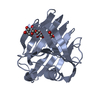
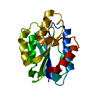
| Title | CATALYSIS AND SPECIFICITY IN ENZYMATIC GLYCOSIDE HYDROLASES: A 2,5B CONFORMATION FOR THE GLYCOSYL-ENZYME INTERMIDIATE REVEALED BY THE STRUCTURE OF THE BACILLUS AGARADHAERENS FAMILY 11 XYLANASE | ||||||||||||
 Components Components | XYLANASE | ||||||||||||
 Keywords Keywords | HYDROLASE / GLYCOSYL HYDROLASE | ||||||||||||
| Function / homology |  Function and homology information Function and homology informationendo-1,4-beta-xylanase / endo-1,4-beta-xylanase activity / xylan catabolic process Similarity search - Function | ||||||||||||
| Biological species |  Bacillus agaradhaerens (bacteria) Bacillus agaradhaerens (bacteria) | ||||||||||||
| Method |  X-RAY DIFFRACTION / MOLECULAR REPLACEMENT / Resolution: 1.78 Å X-RAY DIFFRACTION / MOLECULAR REPLACEMENT / Resolution: 1.78 Å | ||||||||||||
 Authors Authors | Sabini, E. / Sulzenbacher, G. / Dauter, M. / Dauter, Z. / Jorgensen, P.L. / Schulein, M. / Dupont, C. / Davies, G.J. / Wilson, K.S. | ||||||||||||
 Citation Citation | Journal: Chem.Biol. / Year: 1999 Title: Catalysis and specificity in enzymatic glycoside hydrolysis: a 2,5B conformation for the glycosyl-enzyme intermediate revealed by the structure of the Bacillus agaradhaerens family 11 xylanase. Authors: Sabini, E. / Sulzenbacher, G. / Dauter, M. / Dauter, Z. / Jorgensen, P.L. / Schulein, M. / Dupont, C. / Davies, G.J. / Wilson, K.S. | ||||||||||||
| History |
|
- Structure visualization
| Structure viewer | Molecule: MolmilJmol/JSmol |
|---|
- Downloads & links
-Download
| PDBx/mmCIF format | 1qh7.cif.gz | 200.4 KB | Display | PDBx/mmCIF format |
|---|---|---|---|---|
| PDB format | pdb1qh7.ent.gz | 160.2 KB | Display | PDB format |
| PDBx/mmJSON format | 1qh7.json.gz | Tree view | PDBx/mmJSON format | |
| Others |  Other downloads Other downloads |
-Validation report
| Arichive directory | https://data.pdbj.org/pub/pdb/validation_reports/qh/1qh7ftp://data.pdbj.org/pub/pdb/validation_reports/qh/1qh7 | HTTPS FTP |
|---|
-Related structure data


-Links
 PDBj
PDBj
- Assembly
| Deposited unit | 
| ||||||||
|---|---|---|---|---|---|---|---|---|---|
| 1 | 
| ||||||||
| 2 | 
| ||||||||
| Unit cell |
| ||||||||
| Noncrystallographic symmetry (NCS) | NCS oper: (Code: given Matrix: (0.95985, -0.280398, -0.008088), Vector: |
-Components
| #1: Protein | Mass: 23155.547 Da / Num. of mol.: 2 / Fragment: FAMILY 11 XYLANASE CATALYTIC DOMAIN Source method: isolated from a genetically manipulated source Details: B-D-XYLANOPYRANOSIDE PRESENT IN THE ACTIVE SITE / Source: (gene. exp.) Bacillus agaradhaerens (bacteria) / Production host: #2: Sugar |   Type: D-saccharide, beta linking / Mass: 150.130 Da / Num. of mol.: 2 / Source method: obtained synthetically / Formula: C5H10O5 Type: D-saccharide, beta linking / Mass: 150.130 Da / Num. of mol.: 2 / Source method: obtained synthetically / Formula: C5H10O5#3: Water | ChemComp-HOH / |  Mass: 18.015 Da / Num. of mol.: 611 / Source method: isolated from a natural source / Formula: H2O Mass: 18.015 Da / Num. of mol.: 611 / Source method: isolated from a natural source / Formula: H2OHas protein modification | Y | |
|---|
-Experimental details
-Experiment
| Experiment | Method: X-RAY DIFFRACTION / Number of used crystals: 1 |
|---|
- Sample preparation
| Crystal | Density Matthews: 2.4 Å3/Da / Density % sol: 48.6 % | |||||||||||||||||||||||||
|---|---|---|---|---|---|---|---|---|---|---|---|---|---|---|---|---|---|---|---|---|---|---|---|---|---|---|
| Crystal grow | pH: 6.5 / Details: AMMONIUM SULPHATE 30%, MES 0.1M PH 6.5 | |||||||||||||||||||||||||
| Crystal grow | *PLUS pH: 6 / Method: vapor diffusion, hanging drop | |||||||||||||||||||||||||
| Components of the solutions | *PLUS
|
-Data collection
| Diffraction | Mean temperature: 100 K |
|---|---|
| Diffraction source | Source: ROTATING ANODE / Type: RIGAKU RU200 / Wavelength: 1.5418 |
| Detector | Type: MARRESEARCH / Detector: IMAGE PLATE / Date: May 1, 1998 |
| Radiation | Monochromator: NI FILTER / Protocol: SINGLE WAVELENGTH / Monochromatic (M) / Laue (L): M / Scattering type: x-ray |
| Radiation wavelength | Wavelength: 1.5418 Å / Relative weight: 1 |
| Reflection | Resolution: 1.78→54.2 Å / Num. obs: 40782 / % possible obs: 99.1 % / Redundancy: 5.6 % / Biso Wilson estimate: 14.47 Å2 / Rmerge(I) obs: 0.055 / Rsym value: 5.5 / Net I/σ(I): 27.4 |
| Reflection shell | Resolution: 1.78→1.81 Å / Redundancy: 3.2 % / Rmerge(I) obs: 0.162 / Mean I/σ(I) obs: 6.7 / Rsym value: 16.2 / % possible all: 98.3 |
| Reflection shell | *PLUS % possible obs: 98.3 % |
- Processing
| Software |
| ||||||||||||||||||||||||||||||||||||||||||||||||||||||||||||||||||||||||||||||||||||
|---|---|---|---|---|---|---|---|---|---|---|---|---|---|---|---|---|---|---|---|---|---|---|---|---|---|---|---|---|---|---|---|---|---|---|---|---|---|---|---|---|---|---|---|---|---|---|---|---|---|---|---|---|---|---|---|---|---|---|---|---|---|---|---|---|---|---|---|---|---|---|---|---|---|---|---|---|---|---|---|---|---|---|---|---|---|
| Refinement | Method to determine structure: MOLECULAR REPLACEMENT / Resolution: 1.78→20 Å / Cross valid method: THROUGHOUT / σ(F): 0
| ||||||||||||||||||||||||||||||||||||||||||||||||||||||||||||||||||||||||||||||||||||
| Displacement parameters | Biso mean: 16.49 Å2 | ||||||||||||||||||||||||||||||||||||||||||||||||||||||||||||||||||||||||||||||||||||
| Refinement step | Cycle: LAST / Resolution: 1.78→20 Å
| ||||||||||||||||||||||||||||||||||||||||||||||||||||||||||||||||||||||||||||||||||||
| Refine LS restraints |
| ||||||||||||||||||||||||||||||||||||||||||||||||||||||||||||||||||||||||||||||||||||
| Software | *PLUS Name: REFMAC / Classification: refinement | ||||||||||||||||||||||||||||||||||||||||||||||||||||||||||||||||||||||||||||||||||||
| Refinement | *PLUS σ(F): 0 / % reflection Rfree: 5 % / Rfactor obs: 0.117 | ||||||||||||||||||||||||||||||||||||||||||||||||||||||||||||||||||||||||||||||||||||
| Solvent computation | *PLUS | ||||||||||||||||||||||||||||||||||||||||||||||||||||||||||||||||||||||||||||||||||||
| Displacement parameters | *PLUS | ||||||||||||||||||||||||||||||||||||||||||||||||||||||||||||||||||||||||||||||||||||
| Refine LS restraints | *PLUS Type: p_chiral_restr / Dev ideal target: 0.15 |
