 Movie
Movie Controller

[English] 日本語
 Yorodumi
- PDB-1ciz: X-RAY STRUCTURE OF HUMAN STROMELYSIN CATALYTIC DOMAIN COMPLEXES W... -
+ Open data

- Basic information
| Entry | Database: PDB / ID: 1ciz | ||||||
|---|---|---|---|---|---|---|---|
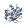
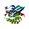
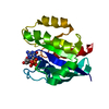
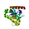
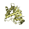
| Title | X-RAY STRUCTURE OF HUMAN STROMELYSIN CATALYTIC DOMAIN COMPLEXES WITH NON-PEPTIDE INHIBITORS: IMPLICATION FOR INHIBITOR SELECTIVITY | ||||||
 Components Components | PROTEIN (STROMELYSIN-1) | ||||||
 Keywords Keywords | METALLOPROTEINASE / MATRIX METALLOPROTEINASE / MMP-3 / NON-PEPTIDE INHIBITOR | ||||||
| Function / homology |  Function and homology information Function and homology informationstromelysin 1 / cellular response to UV-A / regulation of neuroinflammatory response / Assembly of collagen fibrils and other multimeric structures / Activation of Matrix Metalloproteinases / response to amyloid-beta / Collagen degradation / collagen catabolic process / negative regulation of reactive oxygen species metabolic process / extracellular matrix disassembly ...stromelysin 1 / cellular response to UV-A / regulation of neuroinflammatory response / Assembly of collagen fibrils and other multimeric structures / Activation of Matrix Metalloproteinases / response to amyloid-beta / Collagen degradation / collagen catabolic process / negative regulation of reactive oxygen species metabolic process / extracellular matrix disassembly / Degradation of the extracellular matrix / extracellular matrix organization / cellular response to nitric oxide / EGFR Transactivation by Gastrin / regulation of cell migration / negative regulation of phosphatidylinositol 3-kinase/protein kinase B signal transduction / protein catabolic process / cellular response to reactive oxygen species / cellular response to amino acid stimulus / positive regulation of protein-containing complex assembly / metalloendopeptidase activity / metallopeptidase activity / peptidase activity / extracellular matrix / cellular response to lipopolysaccharide / Interleukin-4 and Interleukin-13 signaling / endopeptidase activity / Extra-nuclear estrogen signaling / serine-type endopeptidase activity / innate immune response / mitochondrion / proteolysis / : / extracellular region / zinc ion binding / nucleus / cytosol Similarity search - Function | ||||||
| Biological species |  Homo sapiens (human) Homo sapiens (human) | ||||||
| Method |  X-RAY DIFFRACTION / MOLECULAR REPLACEMENT / Resolution: 1.64 Å X-RAY DIFFRACTION / MOLECULAR REPLACEMENT / Resolution: 1.64 Å | ||||||
 Authors Authors | Pavlovsky, A.G. / Williams, M.G. / Ye, Q.-Z. / Ortwine, D.F. / Purchase II, C.F. / White, A.D. / Dhanaraj, V. / Roth, B.D. / Johnson, L.L. / Hupe, D. ...Pavlovsky, A.G. / Williams, M.G. / Ye, Q.-Z. / Ortwine, D.F. / Purchase II, C.F. / White, A.D. / Dhanaraj, V. / Roth, B.D. / Johnson, L.L. / Hupe, D. / Humblet, C. / Blundell, T.L. | ||||||
 Citation Citation | Journal: Protein Sci. / Year: 1999 Title: X-ray structure of human stromelysin catalytic domain complexed with nonpeptide inhibitors: implications for inhibitor selectivity. Authors: Pavlovsky, A.G. / Williams, M.G. / Ye, Q.Z. / Ortwine, D.F. / Purchase II, C.F. / White, A.D. / Dhanaraj, V. / Roth, B.D. / Johnson, L.L. / Hupe, D. / Humblet, C. / Blundell, T.L. | ||||||
| History |
|
- Structure visualization
| Structure viewer | Molecule: MolmilJmol/JSmol |
|---|
- Downloads & links
-Download
| PDBx/mmCIF format | 1ciz.cif.gz | 54.9 KB | Display | PDBx/mmCIF format |
|---|---|---|---|---|
| PDB format | pdb1ciz.ent.gz | 37.4 KB | Display | PDB format |
| PDBx/mmJSON format | 1ciz.json.gz | Tree view | PDBx/mmJSON format | |
| Others |  Other downloads Other downloads |
-Validation report
| Arichive directory | https://data.pdbj.org/pub/pdb/validation_reports/ci/1cizftp://data.pdbj.org/pub/pdb/validation_reports/ci/1ciz | HTTPS FTP |
|---|
-Related structure data
| Related structure data | 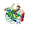 1b8ySC  1caqC  1qiaC  1qicC S: Starting model for refinement C: citing same article ( |
|---|---|
| Similar structure data |


-Links
 PDBj
PDBj


- Assembly
| Deposited unit | 
| ||||||||
|---|---|---|---|---|---|---|---|---|---|
| 1 |
| ||||||||
| 2 | 
| ||||||||
| Unit cell |
| ||||||||
| Components on special symmetry positions |
|
-Components
-Protein , 1 types, 1 molecules A
| #1: Protein | Mass: 18887.029 Da / Num. of mol.: 1 / Fragment: CATALYTIC DOMAIN Source method: isolated from a genetically manipulated source Source: (gene. exp.) Homo sapiens (human) / Cell line: FIBROBLAST / Plasmid: PGEMEX-1 / Production host:  |
|---|
-Non-polymers , 5 types, 154 molecules 


| #2: Chemical |  Mass: 65.409 Da / Num. of mol.: 2 / Source method: obtained synthetically / Formula: Zn Mass: 65.409 Da / Num. of mol.: 2 / Source method: obtained synthetically / Formula: Zn#3: Chemical |  Mass: 40.078 Da / Num. of mol.: 3 / Source method: obtained synthetically / Formula: Ca Mass: 40.078 Da / Num. of mol.: 3 / Source method: obtained synthetically / Formula: Ca#4: Chemical | ChemComp-SO4 / |  Mass: 96.063 Da / Num. of mol.: 1 / Source method: obtained synthetically / Formula: SO4 Mass: 96.063 Da / Num. of mol.: 1 / Source method: obtained synthetically / Formula: SO4#5: Chemical | ChemComp-DPS / |  Mass: 503.613 Da / Num. of mol.: 1 / Source method: obtained synthetically / Formula: C28H29N3O4S Mass: 503.613 Da / Num. of mol.: 1 / Source method: obtained synthetically / Formula: C28H29N3O4S#6: Water | ChemComp-HOH / | Mass: 18.015 Da / Num. of mol.: 147 / Source method: isolated from a natural source / Formula: H2O |
|---|
-Experimental details
-Experiment
| Experiment | Method: X-RAY DIFFRACTION / Number of used crystals: 1 |
|---|
- Sample preparation
| Crystal | Density Matthews: 2.37 Å3/Da / Density % sol: 48.05 % | ||||||||||||||||||||||||||||||||||||||||
|---|---|---|---|---|---|---|---|---|---|---|---|---|---|---|---|---|---|---|---|---|---|---|---|---|---|---|---|---|---|---|---|---|---|---|---|---|---|---|---|---|---|
| Crystal grow | pH: 6.5 / Details: pH 6.5 | ||||||||||||||||||||||||||||||||||||||||
| Crystal grow | *PLUS Method: vapor diffusion | ||||||||||||||||||||||||||||||||||||||||
| Components of the solutions | *PLUS
|
-Data collection
| Diffraction | Mean temperature: 87 K |
|---|---|
| Diffraction source | Source: ROTATING ANODE / Type: RIGAKU RU200 / Wavelength: 1.5418 |
| Detector | Type: MAR scanner 300 mm plate / Detector: IMAGE PLATE / Date: Jan 1, 1997 / Details: SUPPER 8CM/16CM FOCUSING MIRRORS |
| Radiation | Protocol: SINGLE WAVELENGTH / Monochromatic (M) / Laue (L): M / Scattering type: x-ray |
| Radiation wavelength | Wavelength: 1.5418 Å / Relative weight: 1 |
| Reflection | Resolution: 1.64→10 Å / Num. obs: 21098 / % possible obs: 92.3 % / Observed criterion σ(I): 2 / Redundancy: 3.8 % / Rsym value: 0.038 / Net I/σ(I): 22.9 |
| Reflection shell | Resolution: 1.64→1.7 Å / Mean I/σ(I) obs: 11.2 / Rsym value: 0.121 / % possible all: 92.4 |
| Reflection | *PLUS Num. obs: 20986 / Num. measured all: 80181 / Rmerge(I) obs: 0.038 |
| Reflection shell | *PLUS % possible obs: 92.4 % / Rmerge(I) obs: 0.12 |
- Processing
| Software |
| ||||||||||||||||||||||||||||||||||||||||||||||||||||||||||||
|---|---|---|---|---|---|---|---|---|---|---|---|---|---|---|---|---|---|---|---|---|---|---|---|---|---|---|---|---|---|---|---|---|---|---|---|---|---|---|---|---|---|---|---|---|---|---|---|---|---|---|---|---|---|---|---|---|---|---|---|---|---|
| Refinement | Method to determine structure: MOLECULAR REPLACEMENT Starting model: PROTEIN FROM 1B8Y Resolution: 1.64→8 Å / Cross valid method: THROUGHOUT / σ(F): 2
| ||||||||||||||||||||||||||||||||||||||||||||||||||||||||||||
| Refinement step | Cycle: LAST / Resolution: 1.64→8 Å
| ||||||||||||||||||||||||||||||||||||||||||||||||||||||||||||
| Refine LS restraints |
| ||||||||||||||||||||||||||||||||||||||||||||||||||||||||||||
| LS refinement shell | Resolution: 1.6→1.67 Å / Total num. of bins used: 8
| ||||||||||||||||||||||||||||||||||||||||||||||||||||||||||||
| Xplor file | Serial no: 1 / Param file: PARAM19X.PRO / Topol file: TOPH19X.PRO | ||||||||||||||||||||||||||||||||||||||||||||||||||||||||||||
| Software | *PLUS Name: X-PLOR / Version: 3.1 / Classification: refinement | ||||||||||||||||||||||||||||||||||||||||||||||||||||||||||||
| Refinement | *PLUS Lowest resolution: 8 Å / σ(F): 2 / % reflection Rfree: 10 % / Rfactor obs: 0.209 | ||||||||||||||||||||||||||||||||||||||||||||||||||||||||||||
| Solvent computation | *PLUS | ||||||||||||||||||||||||||||||||||||||||||||||||||||||||||||
| Displacement parameters | *PLUS | ||||||||||||||||||||||||||||||||||||||||||||||||||||||||||||
| Refine LS restraints | *PLUS
| ||||||||||||||||||||||||||||||||||||||||||||||||||||||||||||
| LS refinement shell | *PLUS Highest resolution: 1.6 Å / Rfactor Rfree: 0.305 / % reflection Rfree: 11 % / Rfactor Rwork: 0.308 |
