 Movie
Movie Controller

[English] 日本語
 Yorodumi
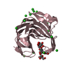
- PDB-1m1u: AN ISOLEUCINE-BASED ALLOSTERIC SWITCH CONTROLS AFFINITY AND SHAPE... -
+ Open data

- Basic information
| Entry | Database: PDB / ID: 1m1u | ||||||
|---|---|---|---|---|---|---|---|
| Title | AN ISOLEUCINE-BASED ALLOSTERIC SWITCH CONTROLS AFFINITY AND SHAPE SHIFTING IN INTEGRIN CD11B A-DOMAIN | ||||||
 Components Components | Integrin alpha-M | ||||||
 Keywords Keywords | CELL ADHESION / INTEGRIN / CELL ADHESION PROTEIN / GLYCOPROTEIN / A-domain / CD11b | ||||||
| Function / homology |  Function and homology information Function and homology informationectodermal cell differentiation / integrin alphaM-beta2 complex / positive regulation of neutrophil degranulation / response to Gram-positive bacterium / response to curcumin / leukocyte adhesion to vascular endothelial cell / positive regulation of microglial cell mediated cytotoxicity / vertebrate eye-specific patterning / complement component C3b binding / complement-mediated synapse pruning ...ectodermal cell differentiation / integrin alphaM-beta2 complex / positive regulation of neutrophil degranulation / response to Gram-positive bacterium / response to curcumin / leukocyte adhesion to vascular endothelial cell / positive regulation of microglial cell mediated cytotoxicity / vertebrate eye-specific patterning / complement component C3b binding / complement-mediated synapse pruning / Toll Like Receptor 4 (TLR4) Cascade / cell-cell adhesion mediated by integrin / complement receptor mediated signaling pathway / integrin complex / heterotypic cell-cell adhesion / cargo receptor activity / phagocytosis, engulfment / forebrain development / negative regulation of dopamine metabolic process / amyloid-beta clearance / tertiary granule membrane / plasma membrane raft / Integrin cell surface interactions / positive regulation of protein targeting to membrane / response to mechanical stimulus / specific granule membrane / positive regulation of superoxide anion generation / heat shock protein binding / receptor-mediated endocytosis / response to ischemia / response to amphetamine / integrin-mediated signaling pathway / cell-matrix adhesion / Cell surface interactions at the vascular wall / cell-cell adhesion / microglial cell activation / integrin binding / response to estradiol / amyloid-beta binding / signaling receptor activity / Interleukin-4 and Interleukin-13 signaling / cell adhesion / external side of plasma membrane / innate immune response / Neutrophil degranulation / cell surface / : / extracellular exosome / metal ion binding / plasma membrane Similarity search - Function | ||||||
| Biological species |  Homo sapiens (human) Homo sapiens (human) | ||||||
| Method |  X-RAY DIFFRACTION / SYNCHROTRON / FOURIER SYNTHESIS / Resolution: 2.3 Å X-RAY DIFFRACTION / SYNCHROTRON / FOURIER SYNTHESIS / Resolution: 2.3 Å | ||||||
 Authors Authors | Xiong, J.-P. / Li, R. / Essafi, M. / Stehle, T. / Arnaout, M.A. | ||||||
 Citation Citation | Journal: J.Biol.Chem. / Year: 2000 Title: An isoleucine-based allosteric switch controls affinity and shape shifting in integrin CD11b A-domain. Authors: Xiong, J.P. / Li, R. / Essafi, M. / Stehle, T. / Arnaout, M.A. | ||||||
| History |
|
- Structure visualization
| Structure viewer | Molecule: MolmilJmol/JSmol |
|---|
- Downloads & links
-Download
| PDBx/mmCIF format | 1m1u.cif.gz | 51.3 KB | Display | PDBx/mmCIF format |
|---|---|---|---|---|
| PDB format | pdb1m1u.ent.gz | 35.7 KB | Display | PDB format |
| PDBx/mmJSON format | 1m1u.json.gz | Tree view | PDBx/mmJSON format | |
| Others |  Other downloads Other downloads |
-Validation report
| Arichive directory | https://data.pdbj.org/pub/pdb/validation_reports/m1/1m1uftp://data.pdbj.org/pub/pdb/validation_reports/m1/1m1u | HTTPS FTP |
|---|
-Related structure data
| Related structure data |  1idoS S: Starting model for refinement |
|---|---|
| Similar structure data |


-Links
 PDBj
PDBj


- Assembly
| Deposited unit | 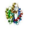
| ||||||||
|---|---|---|---|---|---|---|---|---|---|
| 1 |
| ||||||||
| Unit cell |
|
-Components
| #1: Protein | Mass: 22261.314 Da / Num. of mol.: 1 / Fragment: CD11b A-domain, Residues 123-315 / Mutation: C128S Source method: isolated from a genetically manipulated source Source: (gene. exp.) Homo sapiens (human) / Plasmid: pGEX4T-1 / Production host:  |
|---|---|
| #2: Chemical | ChemComp-CA /   Mass: 40.078 Da / Num. of mol.: 1 / Source method: obtained synthetically / Formula: Ca Mass: 40.078 Da / Num. of mol.: 1 / Source method: obtained synthetically / Formula: Ca |
| #3: Water | ChemComp-HOH /  Mass: 18.015 Da / Num. of mol.: 39 / Source method: isolated from a natural source / Formula: H2O Mass: 18.015 Da / Num. of mol.: 39 / Source method: isolated from a natural source / Formula: H2O |
-Experimental details
-Experiment
| Experiment | Method: X-RAY DIFFRACTION / Number of used crystals: 1 |
|---|
- Sample preparation
| Crystal | Density Matthews: 2.22 Å3/Da / Density % sol: 44.66 % | |||||||||||||||||||||||||||||||||||
|---|---|---|---|---|---|---|---|---|---|---|---|---|---|---|---|---|---|---|---|---|---|---|---|---|---|---|---|---|---|---|---|---|---|---|---|---|
| Crystal grow | Temperature: 298 K / Method: vapor diffusion, hanging drop / pH: 8.2 Details: 15% PEG8K, 0.1M Tris-HCl 8.2, 5mM CaCl2, VAPOR DIFFUSION, HANGING DROP, temperature 298K | |||||||||||||||||||||||||||||||||||
| Crystal grow | *PLUS | |||||||||||||||||||||||||||||||||||
| Components of the solutions | *PLUS
|
-Data collection
| Diffraction | Mean temperature: 100 K |
|---|---|
| Diffraction source | Source: SYNCHROTRON / Site: NSLS  / Beamline: X12B / Wavelength: 0.979 Å / Beamline: X12B / Wavelength: 0.979 Å |
| Detector | Type: ADSC QUANTUM 4 / Detector: CCD / Date: Oct 15, 1999 / Details: mirrors |
| Radiation | Protocol: SINGLE WAVELENGTH / Monochromatic (M) / Laue (L): M / Scattering type: x-ray |
| Radiation wavelength | Wavelength: 0.979 Å / Relative weight: 1 |
| Reflection | Resolution: 2.3→8 Å / Num. obs: 7963 / % possible obs: 91.1 % / Observed criterion σ(I): -2 / Rmerge(I) obs: 0.088 / Rsym value: 0.088 |
| Reflection | *PLUS Num. obs: 7955 / Redundancy: 2.3 % / Rmerge(I) obs: 0.088 |
| Reflection shell | *PLUS % possible obs: 80.8 % / Rmerge(I) obs: 0.28 |
- Processing
| Software |
| ||||||||||||||||||
|---|---|---|---|---|---|---|---|---|---|---|---|---|---|---|---|---|---|---|---|
| Refinement | Method to determine structure: FOURIER SYNTHESIS Starting model: PDB ENTRY 1IDO Resolution: 2.3→8 Å / Cross valid method: througthout / σ(F): 2 / Stereochemistry target values: Engh & Huber
| ||||||||||||||||||
| Refinement step | Cycle: LAST / Resolution: 2.3→8 Å
| ||||||||||||||||||
| Refine LS restraints |
| ||||||||||||||||||
| Refinement | *PLUS % reflection Rfree: 5 % / Rfactor Rfree: 0.248 / Rfactor Rwork: 0.188 | ||||||||||||||||||
| Solvent computation | *PLUS | ||||||||||||||||||
| Displacement parameters | *PLUS |
