 Movie
Movie Controller

[English] 日本語
 Yorodumi
- PDB-1qg3: CRYSTAL STRUCTURE OF A TANDEM PAIR OF FIBRONECTIN TYPE III DOMAIN... -
+ Open data

- Basic information
| Entry | Database: PDB / ID: 1qg3 | ||||||
|---|---|---|---|---|---|---|---|
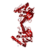
| Title | CRYSTAL STRUCTURE OF A TANDEM PAIR OF FIBRONECTIN TYPE III DOMAINS FROM THE CYTOPLASMIC TAIL OF INTEGRIN ALPHA6 BETA4 | ||||||
 Components Components | PROTEIN (INTEGRIN BETA-4 SUBUNIT) | ||||||
 Keywords Keywords | STRUCTURAL PROTEIN / INTEGRIN / HEMIDESMOSOME / FIBRONECTIN / CARCINOMA | ||||||
| Function / homology |  Function and homology information Function and homology informationType I hemidesmosome assembly / nail development / hemidesmosome assembly / hemidesmosome / peripheral nervous system myelin formation / trophoblast cell migration / skin morphogenesis / Laminin interactions / filopodium assembly / mesodermal cell differentiation ...Type I hemidesmosome assembly / nail development / hemidesmosome assembly / hemidesmosome / peripheral nervous system myelin formation / trophoblast cell migration / skin morphogenesis / Laminin interactions / filopodium assembly / mesodermal cell differentiation / Differentiation of Keratinocytes in Interfollicular Epidermis in Mammalian Skin / Assembly of collagen fibrils and other multimeric structures / integrin complex / cell adhesion mediated by integrin / Syndecan interactions / cell leading edge / basement membrane / basal plasma membrane / cell-matrix adhesion / integrin-mediated signaling pathway / cell motility / cell-cell adhesion / response to wounding / G protein-coupled receptor binding / integrin binding / autophagy / cell junction / cell migration / nuclear membrane / cell adhesion / signaling receptor complex / focal adhesion / nucleolus / cell surface / extracellular exosome / metal ion binding / plasma membrane Similarity search - Function | ||||||
| Biological species |  Homo sapiens (human) Homo sapiens (human) | ||||||
| Method |  X-RAY DIFFRACTION / SYNCHROTRON / MIRAS / Resolution: 2.15 Å X-RAY DIFFRACTION / SYNCHROTRON / MIRAS / Resolution: 2.15 Å | ||||||
 Authors Authors | de Pereda, J.M. / Wiche, G. / Liddington, R.C. | ||||||
 Citation Citation | Journal: EMBO J. / Year: 1999 Title: Crystal structure of a tandem pair of fibronectin type III domains from the cytoplasmic tail of integrin alpha6beta4. Authors: de Pereda, J.M. / Wiche, G. / Liddington, R.C. | ||||||
| History |
|
- Structure visualization
| Structure viewer | Molecule: MolmilJmol/JSmol |
|---|
- Downloads & links
-Download
| PDBx/mmCIF format | 1qg3.cif.gz | 96.8 KB | Display | PDBx/mmCIF format |
|---|---|---|---|---|
| PDB format | pdb1qg3.ent.gz | 74.4 KB | Display | PDB format |
| PDBx/mmJSON format | 1qg3.json.gz | Tree view | PDBx/mmJSON format | |
| Others |  Other downloads Other downloads |
-Validation report
| Arichive directory | https://data.pdbj.org/pub/pdb/validation_reports/qg/1qg3ftp://data.pdbj.org/pub/pdb/validation_reports/qg/1qg3 | HTTPS FTP |
|---|
-Related structure data
| Similar structure data |
|---|


-Links
 PDBj
PDBj


- Assembly
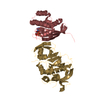
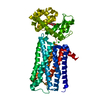
| Deposited unit | 
| |||||||||
|---|---|---|---|---|---|---|---|---|---|---|
| 1 | 
| |||||||||
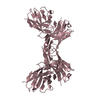
| Unit cell |
| |||||||||
| Components on special symmetry positions |
|
-Components
| #1: Protein | Mass: 21668.338 Da / Num. of mol.: 2 / Fragment: FIRST TANDEM PAIR OF FIBRONECTIN TYPE III DOMAINS Source method: isolated from a genetically manipulated source Source: (gene. exp.) Homo sapiens (human) / Gene: ITGB4 / Species (production host): Escherichia coli / Production host:  #2: Chemical |   Mass: 136.989 Da / Num. of mol.: 2 / Source method: obtained synthetically / Formula: C2H6AsO2 Mass: 136.989 Da / Num. of mol.: 2 / Source method: obtained synthetically / Formula: C2H6AsO2#3: Chemical |   Mass: 96.063 Da / Num. of mol.: 3 / Source method: obtained synthetically / Formula: SO4 Mass: 96.063 Da / Num. of mol.: 3 / Source method: obtained synthetically / Formula: SO4#4: Water | ChemComp-HOH / |  Mass: 18.015 Da / Num. of mol.: 381 / Source method: isolated from a natural source / Formula: H2O Mass: 18.015 Da / Num. of mol.: 381 / Source method: isolated from a natural source / Formula: H2O |
|---|
-Experimental details
-Experiment
| Experiment | Method: X-RAY DIFFRACTION / Number of used crystals: 1 |
|---|
- Sample preparation
| Crystal | Density Matthews: 2.47 Å3/Da / Density % sol: 41 % | ||||||||||||||||||||||||||||||||||||
|---|---|---|---|---|---|---|---|---|---|---|---|---|---|---|---|---|---|---|---|---|---|---|---|---|---|---|---|---|---|---|---|---|---|---|---|---|---|
| Crystal grow | pH: 6.2 Details: 100 MM CACODYLIC ACID/NAOH PH 6.2 200 MM LI2SO4 30% 400 MM POLYETHYLENE GLYCOL | ||||||||||||||||||||||||||||||||||||
| Crystal | *PLUS | ||||||||||||||||||||||||||||||||||||
| Crystal grow | *PLUS Temperature: 4 ℃ / pH: 7.5 / Method: vapor diffusion / Details: used to seeding | ||||||||||||||||||||||||||||||||||||
| Components of the solutions | *PLUS
|
-Data collection
| Diffraction | Mean temperature: 100 K |
|---|---|
| Diffraction source | Source: SYNCHROTRON / Site: APS  / Beamline: 14-BM-C / Wavelength: 1.037 / Beamline: 14-BM-C / Wavelength: 1.037 |
| Detector | Type: ADSC QUANTUM 4 / Detector: CCD / Date: Nov 1, 1998 |
| Radiation | Protocol: SINGLE WAVELENGTH / Monochromatic (M) / Laue (L): M / Scattering type: x-ray |
| Radiation wavelength | Wavelength: 1.037 Å / Relative weight: 1 |
| Reflection | Resolution: 2.15→20 Å / Num. obs: 22644 / % possible obs: 91.5 % / Observed criterion σ(I): 0 / Redundancy: 4.9 % / Biso Wilson estimate: 14.8 Å2 / Rsym value: 0.035 / Net I/σ(I): 34 |
| Reflection shell | Resolution: 2.15→2.21 Å / Mean I/σ(I) obs: 20 / Rsym value: 0.068 / % possible all: 98.9 |
| Reflection | *PLUS Rmerge(I) obs: 0.035 |
| Reflection shell | *PLUS Rmerge(I) obs: 0.068 |
- Processing
| Software |
| ||||||||||||||||||||||||||||||||||||||||||||||||||||||||||||||||||||||||||||||||
|---|---|---|---|---|---|---|---|---|---|---|---|---|---|---|---|---|---|---|---|---|---|---|---|---|---|---|---|---|---|---|---|---|---|---|---|---|---|---|---|---|---|---|---|---|---|---|---|---|---|---|---|---|---|---|---|---|---|---|---|---|---|---|---|---|---|---|---|---|---|---|---|---|---|---|---|---|---|---|---|---|---|
| Refinement | Method to determine structure: MIRAS / Resolution: 2.15→20 Å / Rfactor Rfree error: 0.008 / Data cutoff high rms absF: 2163566.1 / Isotropic thermal model: RESTRAINED / Cross valid method: THROUGHOUT / σ(F): 0
| ||||||||||||||||||||||||||||||||||||||||||||||||||||||||||||||||||||||||||||||||
| Solvent computation | Solvent model: FLAT MODEL / Bsol: 54.49 Å2 / ksol: 0.37 e/Å3 | ||||||||||||||||||||||||||||||||||||||||||||||||||||||||||||||||||||||||||||||||
| Displacement parameters | Biso mean: 26.7 Å2
| ||||||||||||||||||||||||||||||||||||||||||||||||||||||||||||||||||||||||||||||||
| Refine analyze |
| ||||||||||||||||||||||||||||||||||||||||||||||||||||||||||||||||||||||||||||||||
| Refinement step | Cycle: LAST / Resolution: 2.15→20 Å
| ||||||||||||||||||||||||||||||||||||||||||||||||||||||||||||||||||||||||||||||||
| Refine LS restraints |
| ||||||||||||||||||||||||||||||||||||||||||||||||||||||||||||||||||||||||||||||||
| LS refinement shell | Resolution: 2.15→2.28 Å / Rfactor Rfree error: 0.022 / Total num. of bins used: 6
| ||||||||||||||||||||||||||||||||||||||||||||||||||||||||||||||||||||||||||||||||
| Xplor file |
| ||||||||||||||||||||||||||||||||||||||||||||||||||||||||||||||||||||||||||||||||
| Software | *PLUS Name: CNS / Version: 0.5 / Classification: refinement | ||||||||||||||||||||||||||||||||||||||||||||||||||||||||||||||||||||||||||||||||
| Refinement | *PLUS Lowest resolution: 20 Å / σ(F): 0 / % reflection Rfree: 5 % / Rfactor obs: 0.201 | ||||||||||||||||||||||||||||||||||||||||||||||||||||||||||||||||||||||||||||||||
| Solvent computation | *PLUS | ||||||||||||||||||||||||||||||||||||||||||||||||||||||||||||||||||||||||||||||||
| Displacement parameters | *PLUS Biso mean: 26.7 Å2 | ||||||||||||||||||||||||||||||||||||||||||||||||||||||||||||||||||||||||||||||||
| Refine LS restraints | *PLUS
| ||||||||||||||||||||||||||||||||||||||||||||||||||||||||||||||||||||||||||||||||
| LS refinement shell | *PLUS Rfactor Rfree: 0.283 / % reflection Rfree: 4.7 % / Rfactor Rwork: 0.219 |
