 Movie
Movie Controller


+ Open data

- Basic information
| Entry | Database: PDB / ID: 1qdc | |||||||||
|---|---|---|---|---|---|---|---|---|---|---|
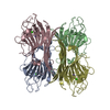
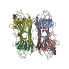
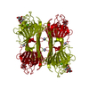
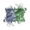
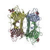
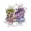
| Title | MAN(APLHA1-6)MAN(ALPHA1-O)METHYL CONCANAVALIN A COMPLEX | |||||||||
 Components Components | PROTEIN (CONCANAVALIN A) | |||||||||
 Keywords Keywords | SUGAR BINDING PROTEIN / PLANT LECTIN / CARBOHYDRATE BINDING / DIMANNOSE / CONCANAVALIN A | |||||||||
| Function / homology |  Function and homology information Function and homology information | |||||||||
| Biological species |   Canavalia ensiformis (jack bean) Canavalia ensiformis (jack bean) | |||||||||
| Method |  X-RAY DIFFRACTION / MOLECULAR REPLACEMENT / Resolution: 2 Å X-RAY DIFFRACTION / MOLECULAR REPLACEMENT / Resolution: 2 Å | |||||||||
 Authors Authors | Bouckaert, J. / Loris, R. / Wyns, L. | |||||||||
 Citation Citation | Journal: J.Biol.Chem. / Year: 1999 Title: The crystal structures of Man(alpha1-3)Man(alpha1-O)Me and Man(alpha1-6)Man(alpha1-O)Me in complex with concanavalin A. Authors: Bouckaert, J. / Hamelryck, T.W. / Wyns, L. / Loris, R. #1: Journal: J.Biol.Chem. / Year: 1996Title: Concanavalin a Crystallized in Complex with the Trisaccharide 3,6-Di-O-(Alpha- D-Mannopyranosyl)-Alpha-D-Mannopyranoside Authors: Loris, R. / Maes, D. / Poortmans, F. / Wyns, L. / Bouckaert, J. #2: Journal: J.Biol.Chem. / Year: 1996Title: Structural Basis of Trimannoside Recognition by Concanavalin A Authors: Naismith, J.H. / Field, R.A. #3: Journal: Biochemistry / Year: 1994Title: Thermodynamics of Lectin-Carbohydrate Interactions. Titration Microcalorimetry Measurements of the Binding of N-Linked Carbohydrates and Ovalbumin to concanavalin A Authors: Mandal, D.K. / Kishore, N. / Brewer, C.F. | |||||||||
| History |
|
- Structure visualization
| Structure viewer | Molecule: MolmilJmol/JSmol |
|---|
- Downloads & links
-Download
| PDBx/mmCIF format | 1qdc.cif.gz | 201.4 KB | Display | PDBx/mmCIF format |
|---|---|---|---|---|
| PDB format | pdb1qdc.ent.gz | 159.2 KB | Display | PDB format |
| PDBx/mmJSON format | 1qdc.json.gz | Tree view | PDBx/mmJSON format | |
| Others |  Other downloads Other downloads |
-Validation report
| Arichive directory | https://data.pdbj.org/pub/pdb/validation_reports/qd/1qdcftp://data.pdbj.org/pub/pdb/validation_reports/qd/1qdc | HTTPS FTP |
|---|
-Related structure data
| Related structure data |  1qdoC  1enqS S: Starting model for refinement C: citing same article ( |
|---|---|
| Similar structure data |


-Links
 PDBj
PDBj

- Assembly
| Deposited unit | 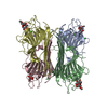
| ||||||||||
|---|---|---|---|---|---|---|---|---|---|---|---|
| 1 |
| ||||||||||
| Unit cell |
| ||||||||||
| Details | THE ASYMMETRIC UNIT CONTAINS A TETRAMER, COMPOSED OF IDENTICAL MONOMERS OF 237 AMINO ACIDS. THE FOLLOWING RESIDUES ARE IN WEAK ELECTRON DENSITY: 118 AND 119, 150 AND 151, 161 AND 162. |
-Components
-Protein / Sugars , 2 types, 8 molecules ABCD
| #1: Protein | Mass: 25622.385 Da / Num. of mol.: 4 / Source method: isolated from a natural source Details: CO-CRYSTALS OF CONCANAVALIN A WITH METHYL-6--O-(ALPHA-D-MANNOPYRANOSYL)-ALPHA- D-MANNOPYRANOSIDE Source: (natural) Canavalia ensiformis (jack bean) / References: UniProt: P55915#2: Polysaccharide | alpha-D-mannopyranose-(1-6)-methyl alpha-D-mannopyranoside Source method: isolated from a genetically manipulated source |
|---|
-Non-polymers , 4 types, 395 molecules 


| #3: Chemical | ChemComp-MN /  Mass: 54.938 Da / Num. of mol.: 4 / Source method: obtained synthetically / Formula: Mn Mass: 54.938 Da / Num. of mol.: 4 / Source method: obtained synthetically / Formula: Mn#4: Chemical | ChemComp-CA /  Mass: 40.078 Da / Num. of mol.: 4 / Source method: obtained synthetically / Formula: Ca Mass: 40.078 Da / Num. of mol.: 4 / Source method: obtained synthetically / Formula: Ca#5: Chemical | ChemComp-CL / |  Mass: 35.453 Da / Num. of mol.: 1 / Source method: obtained synthetically / Formula: Cl Mass: 35.453 Da / Num. of mol.: 1 / Source method: obtained synthetically / Formula: Cl#6: Water | ChemComp-HOH / | Mass: 18.015 Da / Num. of mol.: 386 / Source method: isolated from a natural source / Formula: H2O |
|---|
-Experimental details
-Experiment
| Experiment | Method: X-RAY DIFFRACTION / Number of used crystals: 1 |
|---|
- Sample preparation
| Crystal | Density Matthews: 2.38 Å3/Da / Density % sol: 39.8 % | ||||||||||||||||||||||||||||||||||||||||||||||||
|---|---|---|---|---|---|---|---|---|---|---|---|---|---|---|---|---|---|---|---|---|---|---|---|---|---|---|---|---|---|---|---|---|---|---|---|---|---|---|---|---|---|---|---|---|---|---|---|---|---|
| Crystal grow | Temperature: 291 K / Method: vapor diffusion, hanging drop / pH: 6.8 Details: DROP: 5 MICROLITER 7 MG/ML CON A + 5 MICROLITER 7% JEFFAMINE M-600, 10 % PEG 8000, 50 MM NH4COOH, 5MM MGCL2, 25 MM PHOSPHATE, pH 6.8, VAPOR DIFFUSION, HANGING DROP, temperature 291K | ||||||||||||||||||||||||||||||||||||||||||||||||
| Crystal | *PLUS | ||||||||||||||||||||||||||||||||||||||||||||||||
| Crystal grow | *PLUS | ||||||||||||||||||||||||||||||||||||||||||||||||
| Components of the solutions | *PLUS
|
-Data collection
| Diffraction | Mean temperature: 291 K |
|---|---|
| Diffraction source | Source: ROTATING ANODE / Type: RIGAKU RU200 / Wavelength: 1.5418 |
| Detector | Type: MARRESEARCH / Detector: IMAGE PLATE / Date: Jan 30, 1997 / Details: COLLIMATOR |
| Radiation | Monochromator: GRAPHITE / Protocol: SINGLE WAVELENGTH / Monochromatic (M) / Laue (L): M / Scattering type: x-ray |
| Radiation wavelength | Wavelength: 1.5418 Å / Relative weight: 1 |
| Reflection | Resolution: 2→15 Å / Num. obs: 71100 / % possible obs: 100 % / Observed criterion σ(I): -3 / Redundancy: 7 % / Biso Wilson estimate: 22.1 Å2 / Rmerge(I) obs: 0.082 / Net I/σ(I): 17 |
| Reflection shell | Resolution: 2→2.1 Å / Redundancy: 7 % / Rmerge(I) obs: 0.32 / Mean I/σ(I) obs: 6.3 / % possible all: 99.8 |
| Reflection | *PLUS Num. measured all: 498686 |
| Reflection shell | *PLUS % possible obs: 99.8 % / Rmerge(I) obs: 0.32 |
- Processing
| Software |
| ||||||||||||||||||||||||||||||||||||||||||||||||||||||||||||
|---|---|---|---|---|---|---|---|---|---|---|---|---|---|---|---|---|---|---|---|---|---|---|---|---|---|---|---|---|---|---|---|---|---|---|---|---|---|---|---|---|---|---|---|---|---|---|---|---|---|---|---|---|---|---|---|---|---|---|---|---|---|
| Refinement | Method to determine structure: MOLECULAR REPLACEMENT Starting model: PDB ENTRY 1ENQ Resolution: 2→15 Å / Isotropic thermal model: RESTRAINED / Cross valid method: X-PLOR / σ(F): 0 / σ(I): 0 / Stereochemistry target values: ENGH & HUBER, 1991
| ||||||||||||||||||||||||||||||||||||||||||||||||||||||||||||
| Displacement parameters | Biso mean: 25 Å2 | ||||||||||||||||||||||||||||||||||||||||||||||||||||||||||||
| Refinement step | Cycle: LAST / Resolution: 2→15 Å
| ||||||||||||||||||||||||||||||||||||||||||||||||||||||||||||
| Refine LS restraints |
| ||||||||||||||||||||||||||||||||||||||||||||||||||||||||||||
| LS refinement shell | Resolution: 2→2.09 Å / Total num. of bins used: 8
| ||||||||||||||||||||||||||||||||||||||||||||||||||||||||||||
| Xplor file | Serial no: 1 / Param file: PARHCSDX.PRO / Topol file: TOPHCSDX.PRO | ||||||||||||||||||||||||||||||||||||||||||||||||||||||||||||
| Software | *PLUS Name: X-PLOR / Version: 3.1 / Classification: refinement | ||||||||||||||||||||||||||||||||||||||||||||||||||||||||||||
| Refinement | *PLUS Highest resolution: 2 Å / Lowest resolution: 15 Å / σ(F): 0 / % reflection Rfree: 10 % / Rfactor obs: 0.175 | ||||||||||||||||||||||||||||||||||||||||||||||||||||||||||||
| Solvent computation | *PLUS | ||||||||||||||||||||||||||||||||||||||||||||||||||||||||||||
| Displacement parameters | *PLUS Biso mean: 25 Å2 | ||||||||||||||||||||||||||||||||||||||||||||||||||||||||||||
| Refine LS restraints | *PLUS
| ||||||||||||||||||||||||||||||||||||||||||||||||||||||||||||
| LS refinement shell | *PLUS Highest resolution: 2 Å / Rfactor Rfree: 0.276 / % reflection Rfree: 10 % / Rfactor obs: 0.234 |
