 Movie
Movie Controller


+ Open data

- Basic information
| Entry | Database: PDB / ID: 1azd | ||||||
|---|---|---|---|---|---|---|---|
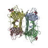
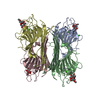
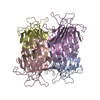
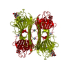
| Title | CONCANAVALIN FROM CANAVALIA BRASILIENSIS | ||||||
 Components Components | CONBR | ||||||
 Keywords Keywords | LECTIN / LEGUME LECTIN | ||||||
| Function / homology |  Function and homology information Function and homology information | ||||||
| Biological species |  Canavalia brasiliensis (Brazilian jackbean) Canavalia brasiliensis (Brazilian jackbean) | ||||||
| Method |  X-RAY DIFFRACTION / DIFFERENCE FOURIER / Resolution: 3 Å X-RAY DIFFRACTION / DIFFERENCE FOURIER / Resolution: 3 Å | ||||||
 Authors Authors | Sanz-Aparicio, J. / Hermoso, J. / Grangeiro, T.B. / Calvete, J.J. / Cavada, B.S. | ||||||
 Citation Citation | Journal: FEBS Lett. / Year: 1997 Title: The crystal structure of Canavalia brasiliensis lectin suggests a correlation between its quaternary conformation and its distinct biological properties from Concanavalin A. Authors: Sanz-Aparicio, J. / Hermoso, J. / Grangeiro, T.B. / Calvete, J.J. / Cavada, B.S. | ||||||
| History |
|
- Structure visualization
| Structure viewer | Molecule: MolmilJmol/JSmol |
|---|
- Downloads & links
-Download
| PDBx/mmCIF format | 1azd.cif.gz | 194.5 KB | Display | PDBx/mmCIF format |
|---|---|---|---|---|
| PDB format | pdb1azd.ent.gz | 157.9 KB | Display | PDB format |
| PDBx/mmJSON format | 1azd.json.gz | Tree view | PDBx/mmJSON format | |
| Others |  Other downloads Other downloads |
-Validation report
| Arichive directory | https://data.pdbj.org/pub/pdb/validation_reports/az/1azdftp://data.pdbj.org/pub/pdb/validation_reports/az/1azd | HTTPS FTP |
|---|
-Related structure data
| Related structure data |  2ctvS S: Starting model for refinement |
|---|---|
| Similar structure data |


-Links
 PDBj
PDBj

- Assembly
| Deposited unit | 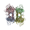
| ||||||||||||||||
|---|---|---|---|---|---|---|---|---|---|---|---|---|---|---|---|---|---|
| 1 |
| ||||||||||||||||
| Unit cell |
| ||||||||||||||||
| Noncrystallographic symmetry (NCS) | NCS oper:
|
-Components
| #1: Protein | Mass: 25592.428 Da / Num. of mol.: 4 / Source method: isolated from a natural source / Details: ISOLATED FROM PLANT Source: (natural) Canavalia brasiliensis (Brazilian jackbean)References: UniProt: P55915 #2: Chemical | ChemComp-CA /   Mass: 40.078 Da / Num. of mol.: 4 / Source method: obtained synthetically / Formula: Ca Mass: 40.078 Da / Num. of mol.: 4 / Source method: obtained synthetically / Formula: Ca#3: Chemical | ChemComp-MN /   Mass: 54.938 Da / Num. of mol.: 4 / Source method: obtained synthetically / Formula: Mn Mass: 54.938 Da / Num. of mol.: 4 / Source method: obtained synthetically / Formula: Mn#4: Water | ChemComp-HOH / |  Mass: 18.015 Da / Num. of mol.: 343 / Source method: isolated from a natural source / Formula: H2O Mass: 18.015 Da / Num. of mol.: 343 / Source method: isolated from a natural source / Formula: H2O |
|---|
-Experimental details
-Experiment
| Experiment | Method: X-RAY DIFFRACTION / Number of used crystals: 1 |
|---|
- Sample preparation
| Crystal | Density Matthews: 2.1 Å3/Da / Density % sol: 40 % | ||||||||||||||||||||||||||||||||||||||||||
|---|---|---|---|---|---|---|---|---|---|---|---|---|---|---|---|---|---|---|---|---|---|---|---|---|---|---|---|---|---|---|---|---|---|---|---|---|---|---|---|---|---|---|---|
| Crystal grow | pH: 6 Details: 2 MICRO-L CBR (10 MG/ML) 2 MICRO-L (11-15% PEG 6000, 0.1 M MES PH 6, pH 6.0 | ||||||||||||||||||||||||||||||||||||||||||
| Crystal | *PLUS | ||||||||||||||||||||||||||||||||||||||||||
| Crystal grow | *PLUS Method: vapor diffusion, hanging drop | ||||||||||||||||||||||||||||||||||||||||||
| Components of the solutions | *PLUS
|
-Data collection
| Diffraction | Mean temperature: 293 K |
|---|---|
| Diffraction source | Source: ROTATING ANODE / Type: ENRAF-NONIUS FR571 / Wavelength: 1.5418 |
| Detector | Type: MARRESEARCH / Detector: IMAGE PLATE / Date: Feb 1, 1996 |
| Radiation | Monochromator: GRAPHITE(002) / Monochromatic (M) / Laue (L): M / Scattering type: x-ray |
| Radiation wavelength | Wavelength: 1.5418 Å / Relative weight: 1 |
| Reflection | Resolution: 2.97→28.4 Å / Num. obs: 16811 / % possible obs: 90 % / Observed criterion σ(I): 0 / Redundancy: 4.1 % / Rmerge(I) obs: 0.15 / Rsym value: 0.15 / Net I/σ(I): 5.7 |
| Reflection shell | Resolution: 2.97→3.06 Å / Redundancy: 4.1 % / Rmerge(I) obs: 0.5 / Mean I/σ(I) obs: 1.5 / Rsym value: 0.5 / % possible all: 88.5 |
| Reflection | *PLUS Num. measured all: 91275 |
- Processing
| Software |
| ||||||||||||||||||||||||||||||||||||||||||||||||||||||||||||
|---|---|---|---|---|---|---|---|---|---|---|---|---|---|---|---|---|---|---|---|---|---|---|---|---|---|---|---|---|---|---|---|---|---|---|---|---|---|---|---|---|---|---|---|---|---|---|---|---|---|---|---|---|---|---|---|---|---|---|---|---|---|
| Refinement | Method to determine structure: DIFFERENCE FOURIER Starting model: PDB ENTRY 2CTV Resolution: 3→8 Å / Data cutoff high absF: 1000000 / Data cutoff low absF: 0.001 / Cross valid method: THROUGHOUT / σ(F): 0
| ||||||||||||||||||||||||||||||||||||||||||||||||||||||||||||
| Displacement parameters | Biso mean: 23.98 Å2 | ||||||||||||||||||||||||||||||||||||||||||||||||||||||||||||
| Refinement step | Cycle: LAST / Resolution: 3→8 Å
| ||||||||||||||||||||||||||||||||||||||||||||||||||||||||||||
| Refine LS restraints |
| ||||||||||||||||||||||||||||||||||||||||||||||||||||||||||||
| LS refinement shell | Resolution: 3→3.13 Å / Total num. of bins used: 8
| ||||||||||||||||||||||||||||||||||||||||||||||||||||||||||||
| Software | *PLUS Name: X-PLOR / Version: 3.843 / Classification: refinement | ||||||||||||||||||||||||||||||||||||||||||||||||||||||||||||
| Refine LS restraints | *PLUS
|
