 Movie
Movie Controller

[English] 日本語
 Yorodumi
- PDB-6vb8: Crystal structure of a lectin from Canavalia brasiliensis seed (C... -
+ Open data

- Basic information
| Entry | Database: PDB / ID: 6vb8 | ||||||
|---|---|---|---|---|---|---|---|
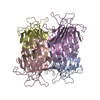
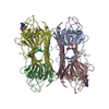
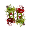
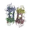
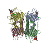
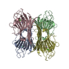
| Title | Crystal structure of a lectin from Canavalia brasiliensis seed (ConBr) complexed with indole-3-acetic acid | ||||||
 Components Components | Concanavalin-Br | ||||||
 Keywords Keywords | SUGAR BINDING PROTEIN / Canavalia brasiliensis / ConBr / IAA / indole-3-acetic acid / auxin / Lectin / ABU / alpha-aminobutyric acid | ||||||
| Function / homology |  Function and homology information Function and homology information | ||||||
| Biological species |  Canavalia brasiliensis (Brazilian jackbean) Canavalia brasiliensis (Brazilian jackbean) | ||||||
| Method |  X-RAY DIFFRACTION / SYNCHROTRON / MOLECULAR REPLACEMENT / Resolution: 2.2 Å X-RAY DIFFRACTION / SYNCHROTRON / MOLECULAR REPLACEMENT / Resolution: 2.2 Å | ||||||
 Authors Authors | Bezerra, E.H.S. / Queiroz, P.P. / da Silva, F.M.S. / Girao, M.S. / Sales, M.V. / Paiva, C.P.S. / Freire, V.N. / Rocha, B.A.M. | ||||||
| Funding support |  Brazil, 1items Brazil, 1items
| ||||||
 Citation Citation | Journal: To Be Published Title: Structural insights into phytohormone interaction to plant lectin ConBr Authors: Bezerra, E.H.S. / Sales, M.V. / Girao, M.S. / da Silva, F.M.S. / Queiroz, P.P. / Paiva, C.P.S. / Freire, V.N. / Rocha, B.A.M. | ||||||
| History |
|
- Structure visualization
| Structure viewer | Molecule: MolmilJmol/JSmol |
|---|
- Downloads & links
-Download
| PDBx/mmCIF format | 6vb8.cif.gz | 107.8 KB | Display | PDBx/mmCIF format |
|---|---|---|---|---|
| PDB format | pdb6vb8.ent.gz | 81 KB | Display | PDB format |
| PDBx/mmJSON format | 6vb8.json.gz | Tree view | PDBx/mmJSON format | |
| Others |  Other downloads Other downloads |
-Validation report
| Arichive directory | https://data.pdbj.org/pub/pdb/validation_reports/vb/6vb8ftp://data.pdbj.org/pub/pdb/validation_reports/vb/6vb8 | HTTPS FTP |
|---|
-Related structure data
| Related structure data |  3ju9S S: Starting model for refinement |
|---|---|
| Similar structure data |


-Links
 PDBj
PDBj


- Assembly
| Deposited unit | 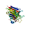
| ||||||||
|---|---|---|---|---|---|---|---|---|---|
| 1 | 
| ||||||||
| Unit cell |
|
-Components
-Protein , 1 types, 1 molecules A
| #1: Protein | Mass: 25592.428 Da / Num. of mol.: 1 / Source method: isolated from a natural source Source: (natural) Canavalia brasiliensis (Brazilian jackbean)References: UniProt: P55915 |
|---|
-Non-polymers , 7 types, 51 molecules 


| #2: Chemical | ChemComp-GOL /  Mass: 92.094 Da / Num. of mol.: 1 / Source method: obtained synthetically / Formula: C3H8O3 Mass: 92.094 Da / Num. of mol.: 1 / Source method: obtained synthetically / Formula: C3H8O3 | ||||||
|---|---|---|---|---|---|---|---|
| #3: Chemical | ChemComp-CA /  Mass: 40.078 Da / Num. of mol.: 1 / Source method: obtained synthetically / Formula: Ca Mass: 40.078 Da / Num. of mol.: 1 / Source method: obtained synthetically / Formula: Ca | ||||||
| #4: Chemical | ChemComp-MN /  Mass: 54.938 Da / Num. of mol.: 1 / Source method: obtained synthetically / Formula: Mn Mass: 54.938 Da / Num. of mol.: 1 / Source method: obtained synthetically / Formula: Mn | ||||||
| #5: Chemical |  Mass: 35.453 Da / Num. of mol.: 2 / Source method: obtained synthetically / Formula: Cl Mass: 35.453 Da / Num. of mol.: 2 / Source method: obtained synthetically / Formula: Cl#6: Chemical | ChemComp-DBB / |  Type: D-peptide linking / Mass: 103.120 Da / Num. of mol.: 1 / Source method: obtained synthetically / Formula: C4H9NO2 Type: D-peptide linking / Mass: 103.120 Da / Num. of mol.: 1 / Source method: obtained synthetically / Formula: C4H9NO2#7: Chemical | ChemComp-IAC / |  Mass: 175.184 Da / Num. of mol.: 1 / Source method: obtained synthetically / Formula: C10H9NO2 / Feature type: SUBJECT OF INVESTIGATION Mass: 175.184 Da / Num. of mol.: 1 / Source method: obtained synthetically / Formula: C10H9NO2 / Feature type: SUBJECT OF INVESTIGATION#8: Water | ChemComp-HOH / | Mass: 18.015 Da / Num. of mol.: 44 / Source method: isolated from a natural source / Formula: H2O |
-Details
| Has ligand of interest | Y |
|---|
-Experimental details
-Experiment
| Experiment | Method: X-RAY DIFFRACTION / Number of used crystals: 1 |
|---|
- Sample preparation
| Crystal | Density Matthews: 2.39 Å3/Da / Density % sol: 48.56 % |
|---|---|
| Crystal grow | Temperature: 298 K / Method: vapor diffusion, hanging drop / pH: 8.5 Details: 1.8 M ammonium sulfate, 0.2 M sodium chloride, 0.1 M HEPES, pH 8.5 |
-Data collection
| Diffraction | Mean temperature: 100 K / Serial crystal experiment: N |
|---|---|
| Diffraction source | Source: SYNCHROTRON / Site: LNLS / Beamline: W01B-MX2 / Wavelength: 1.458 Å |
| Detector | Type: DECTRIS PILATUS 2M / Detector: PIXEL / Date: May 18, 2017 |
| Radiation | Protocol: SINGLE WAVELENGTH / Monochromatic (M) / Laue (L): M / Scattering type: x-ray |
| Radiation wavelength | Wavelength: 1.458 Å / Relative weight: 1 |
| Reflection | Resolution: 2.2→49.69 Å / Num. obs: 12527 / % possible obs: 98.3 % / Redundancy: 11.9 % / CC1/2: 1 / Rmerge(I) obs: 0.046 / Rpim(I) all: 0.013 / Rrim(I) all: 0.048 / Net I/σ(I): 30.4 |
| Reflection shell | Resolution: 2.2→2.32 Å / Redundancy: 8.9 % / Rmerge(I) obs: 0.212 / Mean I/σ(I) obs: 8.9 / Num. unique obs: 1596 / CC1/2: 0.994 / Rpim(I) all: 0.073 / Rrim(I) all: 0.225 / % possible all: 88.8 |
- Processing
| Software |
| ||||||||||||||||||||||||||||||||||||||||||||||||||||||||||||
|---|---|---|---|---|---|---|---|---|---|---|---|---|---|---|---|---|---|---|---|---|---|---|---|---|---|---|---|---|---|---|---|---|---|---|---|---|---|---|---|---|---|---|---|---|---|---|---|---|---|---|---|---|---|---|---|---|---|---|---|---|---|
| Refinement | Method to determine structure: MOLECULAR REPLACEMENT Starting model: 3JU9 Resolution: 2.2→49.69 Å / Cor.coef. Fo:Fc: 0.948 / Cor.coef. Fo:Fc free: 0.93 / SU B: 13.134 / SU ML: 0.159 / Cross valid method: FREE R-VALUE / σ(F): 0 / ESU R: 0.289 / ESU R Free: 0.21 Details: U VALUES : WITH TLS ADDED HYDROGENS HAVE BEEN ADDED IN THE RIDING POSITIONS
| ||||||||||||||||||||||||||||||||||||||||||||||||||||||||||||
| Solvent computation | Ion probe radii: 1 Å / Shrinkage radii: 1 Å / VDW probe radii: 1.2 Å | ||||||||||||||||||||||||||||||||||||||||||||||||||||||||||||
| Displacement parameters | Biso max: 84.67 Å2 / Biso mean: 45.966 Å2 / Biso min: 28.47 Å2
| ||||||||||||||||||||||||||||||||||||||||||||||||||||||||||||
| Refinement step | Cycle: final / Resolution: 2.2→49.69 Å
| ||||||||||||||||||||||||||||||||||||||||||||||||||||||||||||
| Refine LS restraints |
| ||||||||||||||||||||||||||||||||||||||||||||||||||||||||||||
| LS refinement shell | Resolution: 2.2→2.257 Å / Rfactor Rfree error: 0 / Total num. of bins used: 20
| ||||||||||||||||||||||||||||||||||||||||||||||||||||||||||||
| Refinement TLS params. | Method: refined / Origin x: 14.386 Å / Origin y: 14.256 Å / Origin z: 10.956 Å
|
