Mass: 18.015 Da / Num. of mol.: 244 / Source method: isolated from a natural source / Formula: H2O
-
Experimental details
-
Experiment
Experiment
Method: X-RAY DIFFRACTION / Number of used crystals: 1
-
Sample preparation
Crystal
Density Matthews: 2.35 Å3/Da / Density % sol: 47.7 %
Crystal grow
Temperature: 300 K / Method: vapor diffusion, hanging drop / pH: 7.2 Details: each 0.2 mM ConA in 20 mM HEPES, 100 mM NaCl, 5 mM CaCl2, 5 mM MnCl2 pH 7.2
Protocol: SINGLE WAVELENGTH / Monochromatic (M) / Laue (L): M / Scattering type: x-ray
Radiation wavelength
Wavelength: 0.9716 Å / Relative weight: 1
Reflection
Resolution: 2.59→30 Å / % possible obs: 94.4 % / Redundancy: 3.8 % / Net I/σ(I): 18
-
Processing
Software
Name
Version
Classification
REFMAC
5.8.0131
refinement
HKL-2000
datareduction
HKL-2000
datascaling
PHASER
phasing
Refinement
Resolution: 2.59→30 Å / Cor.coef. Fo:Fc: 0.91 / Cor.coef. Fo:Fc free: 0.825 / SU B: 17.086 / SU ML: 0.376 / Cross valid method: THROUGHOUT / ESU R: 1.905 / ESU R Free: 0.475 / Stereochemistry target values: MAXIMUM LIKELIHOOD / Details: HYDROGENS HAVE BEEN ADDED IN THE RIDING POSITIONS
Rfactor
Num. reflection
% reflection
Selection details
Rfree
0.37192
1619
5 %
RANDOM
Rwork
0.27832
-
-
-
obs
0.28303
30451
94.2 %
-
Solvent computation
Ion probe radii: 0.8 Å / Shrinkage radii: 0.8 Å / VDW probe radii: 1.2 Å / Solvent model: MASK
 Movie
Movie Controller


 Open data
 Basic information
 Components
 Keywords
 Function and homology information
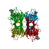
 Canavalia ensiformis (jack bean)
 X-RAY DIFFRACTION /
  Authors
 Citation
 Structure visualization
 Downloads & links
 Other downloads


 PDBj
PDBj


 Assembly
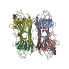


 Type: D-saccharide, alpha linking / Mass: 180.156 Da / Num. of mol.: 2


 Mass: 54.938 Da / Num. of mol.: 4 / Source method: obtained synthetically / Formula: Mn
 Mass: 40.078 Da / Num. of mol.: 6 / Source method: obtained synthetically / Formula: Ca
 Mass: 99.091 Da / Num. of mol.: 2
 Sample preparation
 / Beamline: BL17U1 / Wavelength: 0.9716 Å
 Processing