 Movie
Movie Controller

[English] 日本語
 Yorodumi
- PDB-2je9: CRYSTAL STRUCTURE OF RECOMBINANT DIOCLEA GRANDIFLORA LECTIN COMPL... -
+ Open data

- Basic information
| Entry | Database: PDB / ID: 2je9 | ||||||
|---|---|---|---|---|---|---|---|
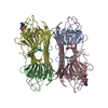
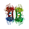
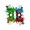
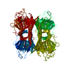
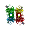
| Title | CRYSTAL STRUCTURE OF RECOMBINANT DIOCLEA GRANDIFLORA LECTIN COMPLEXED WITH 5-BROMO-4-CHLORO-3-INDOLYL-A-D-MANNOSE | ||||||
 Components Components | LECTIN ALPHA CHAIN | ||||||
 Keywords Keywords | SUGAR BINDING PROTEIN / METAL-BINDING / CARBOHYDRATE BINDING PROTEIN / CONA-LIKE / LEGUME LECTIN / SUGAR-BINDING PROTEIN | ||||||
| Function / homology |  Function and homology information Function and homology informationD-mannose binding / toxin activity / carbohydrate binding / metal ion binding / identical protein binding Similarity search - Function | ||||||
| Biological species |  DIOCLEA GRANDIFLORA (mucana) DIOCLEA GRANDIFLORA (mucana) | ||||||
| Method |  X-RAY DIFFRACTION / SYNCHROTRON / MOLECULAR REPLACEMENT / Resolution: 2.1 Å X-RAY DIFFRACTION / SYNCHROTRON / MOLECULAR REPLACEMENT / Resolution: 2.1 Å | ||||||
 Authors Authors | Nagano, C.S. / Sanz, L. / Cavada, B.S. / Calvete, J.J. | ||||||
 Citation Citation | Journal: Biochem.J. / Year: 2008 Title: Insights Into the Structural Basis of the Ph- Dependent Dimer-Tetramer Equilibrium Through Crystallographic Analysis of Recombinant Diocleinae Lectins. Authors: Nagano, C.S. / Calvete, J.J. / Barettino, D. / Perez, A. / Cavada, B.S. / Sanz, L. | ||||||
| History |
| ||||||
| Remark 700 | SHEET DETERMINATION METHOD: AUTHOR PROVIDED. |
- Structure visualization
| Structure viewer | Molecule: MolmilJmol/JSmol |
|---|
- Downloads & links
-Download
| PDBx/mmCIF format | 2je9.cif.gz | 205.6 KB | Display | PDBx/mmCIF format |
|---|---|---|---|---|
| PDB format | pdb2je9.ent.gz | 165.4 KB | Display | PDB format |
| PDBx/mmJSON format | 2je9.json.gz | Tree view | PDBx/mmJSON format | |
| Others |  Other downloads Other downloads |
-Validation report
| Arichive directory | https://data.pdbj.org/pub/pdb/validation_reports/je/2je9ftp://data.pdbj.org/pub/pdb/validation_reports/je/2je9 | HTTPS FTP |
|---|
-Related structure data
| Related structure data |  2jdzC 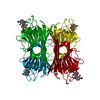 2je7C 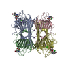 2jecC 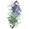 1dglS S: Starting model for refinement C: citing same article ( |
|---|---|
| Similar structure data |


-Links
 PDBj
PDBj

- Assembly
| Deposited unit | 
| ||||||||
|---|---|---|---|---|---|---|---|---|---|
| 1 |
| ||||||||
| Unit cell |
|
-Components
-Protein / Sugars , 2 types, 8 molecules ABCD
| #1: Protein | Mass: 25833.648 Da / Num. of mol.: 4 Source method: isolated from a genetically manipulated source Source: (gene. exp.) DIOCLEA GRANDIFLORA (mucana) / Production host:  #4: Sugar | ChemComp-XMM /  Type: D-saccharide / Mass: 408.629 Da / Num. of mol.: 4 Type: D-saccharide / Mass: 408.629 Da / Num. of mol.: 4Source method: isolated from a genetically manipulated source Formula: C14H15BrClNO6 |
|---|
-Non-polymers , 4 types, 456 molecules 


| #2: Chemical | ChemComp-CA /  Mass: 40.078 Da / Num. of mol.: 4 / Source method: obtained synthetically / Formula: Ca Mass: 40.078 Da / Num. of mol.: 4 / Source method: obtained synthetically / Formula: Ca#3: Chemical | ChemComp-MN /  Mass: 54.938 Da / Num. of mol.: 4 / Source method: obtained synthetically / Formula: Mn Mass: 54.938 Da / Num. of mol.: 4 / Source method: obtained synthetically / Formula: Mn#5: Chemical | ChemComp-SO4 /  Mass: 96.063 Da / Num. of mol.: 7 / Source method: obtained synthetically / Formula: SO4 Mass: 96.063 Da / Num. of mol.: 7 / Source method: obtained synthetically / Formula: SO4#6: Water | ChemComp-HOH / | Mass: 18.015 Da / Num. of mol.: 441 / Source method: isolated from a natural source / Formula: H2O |
|---|
-Details
| Sequence details | THE AMINO ACID SEQUENCES WERE OBTAINED FROM THE DEDUCED DNA. THESE SEQUENCES WERE CONFIRMED BY DNA ...THE AMINO ACID SEQUENCES WERE OBTAINED FROM THE DEDUCED DNA. THESE SEQUENCES WERE CONFIRMED BY DNA SEQUENCE OF ALL CLONES. THE INSERTIONS |
|---|
-Experimental details
-Experiment
| Experiment | Method: X-RAY DIFFRACTION / Number of used crystals: 1 |
|---|
- Sample preparation
| Crystal | Density Matthews: 2.62 Å3/Da / Density % sol: 53.02 % / Description: NONE |
|---|---|
| Crystal grow | Method: vapor diffusion, hanging drop / pH: 7.5 Details: CRYSTALS WERE GROWN BY THE HANGING DROP VAPOUR DIFFUSION METHOD USING 15% PEG 4000, 0.1M HEPES, PH 7.5 AND 0.1M LITHIUM SULFATE |
-Data collection
| Diffraction | Mean temperature: 100 K |
|---|---|
| Diffraction source | Source: SYNCHROTRON / Site: ESRF  / Beamline: ID23-2 / Wavelength: 0.873 / Beamline: ID23-2 / Wavelength: 0.873 |
| Detector | Type: MARRESEARCH / Detector: CCD / Date: Mar 21, 2006 |
| Radiation | Protocol: SINGLE WAVELENGTH / Monochromatic (M) / Laue (L): M / Scattering type: x-ray |
| Radiation wavelength | Wavelength: 0.873 Å / Relative weight: 1 |
| Reflection | Resolution: 2.1→72.17 Å / Num. obs: 63973 / % possible obs: 99.9 % / Observed criterion σ(I): 2 / Redundancy: 4.6 % / Rmerge(I) obs: 0.08 / Net I/σ(I): 6.9 |
| Reflection shell | Resolution: 2.1→2.21 Å / Redundancy: 4 % / Rmerge(I) obs: 0.3 / Mean I/σ(I) obs: 2.1 / % possible all: 99.7 |
- Processing
| Software |
| ||||||||||||||||||||||||||||||||||||||||||||||||||||||||||||||||||||||||||||||||||||||||||||||||||||||||||||||||||||||||||||||||||||||||||||||||||||||||||||||||||||||||||||||||||||||
|---|---|---|---|---|---|---|---|---|---|---|---|---|---|---|---|---|---|---|---|---|---|---|---|---|---|---|---|---|---|---|---|---|---|---|---|---|---|---|---|---|---|---|---|---|---|---|---|---|---|---|---|---|---|---|---|---|---|---|---|---|---|---|---|---|---|---|---|---|---|---|---|---|---|---|---|---|---|---|---|---|---|---|---|---|---|---|---|---|---|---|---|---|---|---|---|---|---|---|---|---|---|---|---|---|---|---|---|---|---|---|---|---|---|---|---|---|---|---|---|---|---|---|---|---|---|---|---|---|---|---|---|---|---|---|---|---|---|---|---|---|---|---|---|---|---|---|---|---|---|---|---|---|---|---|---|---|---|---|---|---|---|---|---|---|---|---|---|---|---|---|---|---|---|---|---|---|---|---|---|---|---|---|---|
| Refinement | Method to determine structure: MOLECULAR REPLACEMENT Starting model: PDB ENTRY 1DGL Resolution: 2.1→87.71 Å / Cor.coef. Fo:Fc: 0.944 / Cor.coef. Fo:Fc free: 0.913 / SU B: 4.569 / SU ML: 0.124 / Cross valid method: THROUGHOUT / ESU R: 0.21 / ESU R Free: 0.185 / Stereochemistry target values: MAXIMUM LIKELIHOOD / Details: HYDROGENS HAVE BEEN ADDED IN THE RIDING POSITIONS
| ||||||||||||||||||||||||||||||||||||||||||||||||||||||||||||||||||||||||||||||||||||||||||||||||||||||||||||||||||||||||||||||||||||||||||||||||||||||||||||||||||||||||||||||||||||||
| Solvent computation | Ion probe radii: 0.8 Å / Shrinkage radii: 0.8 Å / VDW probe radii: 1.4 Å / Solvent model: MASK | ||||||||||||||||||||||||||||||||||||||||||||||||||||||||||||||||||||||||||||||||||||||||||||||||||||||||||||||||||||||||||||||||||||||||||||||||||||||||||||||||||||||||||||||||||||||
| Displacement parameters | Biso mean: 24.47 Å2
| ||||||||||||||||||||||||||||||||||||||||||||||||||||||||||||||||||||||||||||||||||||||||||||||||||||||||||||||||||||||||||||||||||||||||||||||||||||||||||||||||||||||||||||||||||||||
| Refinement step | Cycle: LAST / Resolution: 2.1→87.71 Å
| ||||||||||||||||||||||||||||||||||||||||||||||||||||||||||||||||||||||||||||||||||||||||||||||||||||||||||||||||||||||||||||||||||||||||||||||||||||||||||||||||||||||||||||||||||||||
| Refine LS restraints |
|