 Movie
Movie Controller


+ Open data

- Basic information
| Entry | Database: PDB / ID: 2ef6 | |||||||||
|---|---|---|---|---|---|---|---|---|---|---|
| Title | Canavalia gladiata lectin complexed with Man1-3Man-OMe | |||||||||
 Components Components | Concanavalin A | |||||||||
 Keywords Keywords | PLANT PROTEIN / Canavalia gladiata / lectin / seeds / dimannosides | |||||||||
| Function / homology |  Function and homology information Function and homology informationregulation of defense response to virus / monosaccharide binding / D-mannose binding / positive regulation of cell division / positive regulation of mitotic nuclear division / negative regulation of DNA-templated transcription / metal ion binding Similarity search - Function | |||||||||
| Biological species |  Canavalia gladiata (sword bean) Canavalia gladiata (sword bean) | |||||||||
| Method |  X-RAY DIFFRACTION / SYNCHROTRON / MOLECULAR REPLACEMENT / Resolution: 2.1 Å X-RAY DIFFRACTION / SYNCHROTRON / MOLECULAR REPLACEMENT / Resolution: 2.1 Å | |||||||||
 Authors Authors | Moreno, F.B.M.B. / Bezerra, G.A. / Oliveira, T.M. / de Souza, E.P. / da Rocha, B.A.M. / Benevides, R.G. / Delatorre, P. / Cavada, B.S. / de Azevedo Jr., W.F. | |||||||||
 Citation Citation | Journal: J.Struct.Biol. / Year: 2007 Title: Structural analysis of Canavalia maritima and Canavalia gladiata lectins complexed with different dimannosides: New insights into the understanding of the structure-biological activity ...Title: Structural analysis of Canavalia maritima and Canavalia gladiata lectins complexed with different dimannosides: New insights into the understanding of the structure-biological activity relationship in legume lectins Authors: Bezerra, G.A. / Oliveira, T.M. / Moreno, F.B.M.B. / de Souza, E.P. / da Rocha, B.A.M. / Benevides, R.G. / Delatorre, P. / de Azevedo Jr., W.F. / Cavada, B.S. | |||||||||
| History |
|
- Structure visualization
| Structure viewer | Molecule: MolmilJmol/JSmol |
|---|
- Downloads & links
-Download
| PDBx/mmCIF format | 2ef6.cif.gz | 200 KB | Display | PDBx/mmCIF format |
|---|---|---|---|---|
| PDB format | pdb2ef6.ent.gz | 158.2 KB | Display | PDB format |
| PDBx/mmJSON format | 2ef6.json.gz | Tree view | PDBx/mmJSON format | |
| Others |  Other downloads Other downloads |
-Validation report
| Arichive directory | https://data.pdbj.org/pub/pdb/validation_reports/ef/2ef6ftp://data.pdbj.org/pub/pdb/validation_reports/ef/2ef6 | HTTPS FTP |
|---|
-Related structure data
| Related structure data | 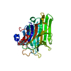 2ovuC 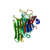 2ow4C 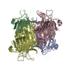 2p2kC  2p34C  2p37C  1wuvS S: Starting model for refinement C: citing same article ( |
|---|---|
| Similar structure data |


-Links
 PDBj
PDBj

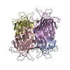
- Assembly
| Deposited unit | 
| ||||||||
|---|---|---|---|---|---|---|---|---|---|
| 1 |
| ||||||||
| Unit cell |
|
-Components
| #1: Protein | Mass: 25563.322 Da / Num. of mol.: 4 / Source method: isolated from a natural source / Source: (natural) Canavalia gladiata (sword bean) / Tissue: seed / References: UniProt: P14894#2: Polysaccharide | alpha-D-mannopyranose-(1-3)-methyl alpha-D-mannopyranoside Source method: isolated from a genetically manipulated source #3: Chemical | ChemComp-CA /   Mass: 40.078 Da / Num. of mol.: 4 / Source method: obtained synthetically / Formula: Ca Mass: 40.078 Da / Num. of mol.: 4 / Source method: obtained synthetically / Formula: Ca#4: Chemical | ChemComp-MN /   Mass: 54.938 Da / Num. of mol.: 4 / Source method: obtained synthetically / Formula: Mn Mass: 54.938 Da / Num. of mol.: 4 / Source method: obtained synthetically / Formula: Mn#5: Water | ChemComp-HOH / |  Mass: 18.015 Da / Num. of mol.: 407 / Source method: isolated from a natural source / Formula: H2O Mass: 18.015 Da / Num. of mol.: 407 / Source method: isolated from a natural source / Formula: H2O |
|---|
-Experimental details
-Experiment
| Experiment | Method: X-RAY DIFFRACTION / Number of used crystals: 1 |
|---|
- Sample preparation
| Crystal | Density Matthews: 2.19 Å3/Da / Density % sol: 43.88 % |
|---|---|
| Crystal grow | Temperature: 293 K / Method: vapor diffusion, hanging drop / pH: 4.5 Details: Sodium Formate, pH 4.5, VAPOR DIFFUSION, HANGING DROP, temperature 293K |
-Data collection
| Diffraction | Mean temperature: 100 K |
|---|---|
| Diffraction source | Source: SYNCHROTRON / Site: LNLS  / Beamline: D03B-MX1 / Wavelength: 1.43 Å / Beamline: D03B-MX1 / Wavelength: 1.43 Å |
| Detector | Type: MAR CCD 130 mm / Detector: CCD / Date: Apr 1, 2006 |
| Radiation | Protocol: SINGLE WAVELENGTH / Monochromatic (M) / Laue (L): M / Scattering type: x-ray |
| Radiation wavelength | Wavelength: 1.43 Å / Relative weight: 1 |
| Reflection | Resolution: 2.07→40.29 Å / Num. obs: 48437 / Redundancy: 2.5 % / Rmerge(I) obs: 0.04 / Rsym value: 0.04 / Net I/σ(I): 10.7 |
| Reflection shell | Highest resolution: 2.07 Å / Rmerge(I) obs: 0.452 / Mean I/σ(I) obs: 2 / Rsym value: 0.452 |
- Processing
| Software |
| |||||||||||||||||||||||||||||||||||||||||||||||||||||||||||||||||||||||||||||||||||||||||||||||
|---|---|---|---|---|---|---|---|---|---|---|---|---|---|---|---|---|---|---|---|---|---|---|---|---|---|---|---|---|---|---|---|---|---|---|---|---|---|---|---|---|---|---|---|---|---|---|---|---|---|---|---|---|---|---|---|---|---|---|---|---|---|---|---|---|---|---|---|---|---|---|---|---|---|---|---|---|---|---|---|---|---|---|---|---|---|---|---|---|---|---|---|---|---|---|---|---|
| Refinement | Method to determine structure: MOLECULAR REPLACEMENT Starting model: 1WUV Resolution: 2.1→33.92 Å / Cor.coef. Fo:Fc: 0.945 / Cor.coef. Fo:Fc free: 0.915 / SU B: 5.188 / SU ML: 0.142 / Cross valid method: THROUGHOUT / ESU R: 0.272 / ESU R Free: 0.211 / Stereochemistry target values: MAXIMUM LIKELIHOOD / Details: HYDROGENS HAVE BEEN ADDED IN THE RIDING POSITIONS
| |||||||||||||||||||||||||||||||||||||||||||||||||||||||||||||||||||||||||||||||||||||||||||||||
| Solvent computation | Ion probe radii: 0.8 Å / Shrinkage radii: 0.8 Å / VDW probe radii: 1.4 Å / Solvent model: MASK | |||||||||||||||||||||||||||||||||||||||||||||||||||||||||||||||||||||||||||||||||||||||||||||||
| Displacement parameters | Biso mean: 22.923 Å2
| |||||||||||||||||||||||||||||||||||||||||||||||||||||||||||||||||||||||||||||||||||||||||||||||
| Refinement step | Cycle: LAST / Resolution: 2.1→33.92 Å
| |||||||||||||||||||||||||||||||||||||||||||||||||||||||||||||||||||||||||||||||||||||||||||||||
| Refine LS restraints |
| |||||||||||||||||||||||||||||||||||||||||||||||||||||||||||||||||||||||||||||||||||||||||||||||
| LS refinement shell | Resolution: 2.1→2.155 Å / Total num. of bins used: 20
|
