 Movie
Movie Controller


+ Open data

- Basic information
| Entry | Database: PDB / ID: 1h9p | ||||||
|---|---|---|---|---|---|---|---|
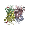
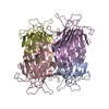
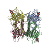
| Title | Crystal Structure of Dioclea guianensis Seed Lectin | ||||||
 Components Components | LECTIN ALPHA CHAIN | ||||||
 Keywords Keywords | LECTIN / LEGUME LECTIN OLIGOMERISATION | ||||||
| Function / homology |  Function and homology information Function and homology informationD-mannose binding / toxin activity / carbohydrate binding / metal ion binding Similarity search - Function | ||||||
| Biological species |  DIOCLEA GUIANENSIS (plant) DIOCLEA GUIANENSIS (plant) | ||||||
| Method |  X-RAY DIFFRACTION / SYNCHROTRON / MOLECULAR REPLACEMENT / Resolution: 2 Å X-RAY DIFFRACTION / SYNCHROTRON / MOLECULAR REPLACEMENT / Resolution: 2 Å | ||||||
 Authors Authors | Romero, A. / Wah, D.A. / Gallego Del sol, F. / Cavada, B.S. / Ramos, M.V. / Grangeiro, T.B. / Sampaio, A.H. / Calvete, J.J. | ||||||
 Citation Citation | Journal: J.Mol.Biol. / Year: 2001 Title: Crystal Structure of Native and Cd/Cd-Substituted Dioclea Guianensis Seed Lectin. A Novel Manganese-Binding Site and Structural Basis of Dimer-Tetramer Association Authors: Wah, D.A. / Romero, A. / Gallego, F. / Cavada, B.S. / Ramos, M.V. / Grangeiro, T.B. / Sampaio, A.H. / Calvete, J.J. #1: Journal: Biochim.Biophys.Acta / Year: 1999 Title: Molecular Characterization and Crystallization of Diocleinae Lectins Authors: Calvete, J.J. / Thole, H.H. / Raida, M. / Urbanke, C. / Romero, A. / Grangeiro, T.B. / Ramos, M.V. / Almeida, I.M. / Guimaraes, F.N. / Cavada, B.S. | ||||||
| History |
|
- Structure visualization
| Structure viewer | Molecule: MolmilJmol/JSmol |
|---|
- Downloads & links
-Download
| PDBx/mmCIF format | 1h9p.cif.gz | 63.2 KB | Display | PDBx/mmCIF format |
|---|---|---|---|---|
| PDB format | pdb1h9p.ent.gz | 45.3 KB | Display | PDB format |
| PDBx/mmJSON format | 1h9p.json.gz | Tree view | PDBx/mmJSON format | |
| Others |  Other downloads Other downloads |
-Validation report
| Arichive directory | https://data.pdbj.org/pub/pdb/validation_reports/h9/1h9pftp://data.pdbj.org/pub/pdb/validation_reports/h9/1h9p | HTTPS FTP |
|---|
-Related structure data
| Related structure data |  1h9wC  1cvnS S: Starting model for refinement C: citing same article ( |
|---|---|
| Similar structure data |


-Links
 PDBj
PDBj

- Assembly
| Deposited unit | 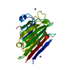
| ||||||||||||||||||||||||
|---|---|---|---|---|---|---|---|---|---|---|---|---|---|---|---|---|---|---|---|---|---|---|---|---|---|
| 1 | 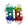
| ||||||||||||||||||||||||
| Unit cell |
| ||||||||||||||||||||||||
| Components on special symmetry positions |
|
-Components
| #1: Protein | Mass: 25451.080 Da / Num. of mol.: 1 / Source method: isolated from a natural source / Source: (natural) DIOCLEA GUIANENSIS (plant) / Variant: LASIOPHYLLA / Tissue: SEED / References: UniProt: P81637 | ||||||
|---|---|---|---|---|---|---|---|
| #2: Chemical |   Mass: 112.411 Da / Num. of mol.: 3 / Source method: obtained synthetically / Formula: Cd Mass: 112.411 Da / Num. of mol.: 3 / Source method: obtained synthetically / Formula: Cd#3: Chemical |   Mass: 54.938 Da / Num. of mol.: 3 / Source method: obtained synthetically / Formula: Mn Mass: 54.938 Da / Num. of mol.: 3 / Source method: obtained synthetically / Formula: Mn#4: Water | ChemComp-HOH / |  Mass: 18.015 Da / Num. of mol.: 128 / Source method: isolated from a natural source / Formula: H2O Mass: 18.015 Da / Num. of mol.: 128 / Source method: isolated from a natural source / Formula: H2OCompound details | D-MANNOSE/D-GLUCOSE-BINDING LECTIN. REQUIRES CA2+ AND MN2+ IONS FOR FULL ACTIVITY. | |
-Experimental details
-Experiment
| Experiment | Method: X-RAY DIFFRACTION / Number of used crystals: 1 |
|---|
- Sample preparation
| Crystal | Density Matthews: 2.17 Å3/Da / Density % sol: 42.96 % | ||||||||||||||||||||||||||||||||||||||||||||||||
|---|---|---|---|---|---|---|---|---|---|---|---|---|---|---|---|---|---|---|---|---|---|---|---|---|---|---|---|---|---|---|---|---|---|---|---|---|---|---|---|---|---|---|---|---|---|---|---|---|---|
| Crystal grow | Method: vapor diffusion, hanging drop / pH: 6.4 Details: CRYSTALS WERE GROWN BY THE HANGING DROP VAPOUR DIFFUSION METHOD USING 30% PEG 400, 0.1M MES, PH 6.4 AND 0.1M CDCL2. | ||||||||||||||||||||||||||||||||||||||||||||||||
| Crystal grow | *PLUS pH: 6.2 / Method: vapor diffusion, hanging drop | ||||||||||||||||||||||||||||||||||||||||||||||||
| Components of the solutions | *PLUS
|
-Data collection
| Diffraction | Mean temperature: 100 K |
|---|---|
| Diffraction source | Source: SYNCHROTRON / Site: EMBL/DESY, HAMBURG  / Beamline: BW7B / Wavelength: 0.8423 / Beamline: BW7B / Wavelength: 0.8423 |
| Detector | Type: MARRESEARCH / Detector: IMAGE PLATE / Date: Oct 15, 2000 / Details: BENT MIRROR |
| Radiation | Monochromator: TRIANGULAR MONOCHROMATOR / Protocol: SINGLE WAVELENGTH / Monochromatic (M) / Laue (L): M / Scattering type: x-ray |
| Radiation wavelength | Wavelength: 0.8423 Å / Relative weight: 1 |
| Reflection | Resolution: 2→35 Å / Num. obs: 15334 / % possible obs: 99.4 % / Redundancy: 6.4 % / Biso Wilson estimate: 15.9 Å2 / Rmerge(I) obs: 0.045 / Net I/σ(I): 12 |
| Reflection shell | Resolution: 2→2.11 Å / Redundancy: 6.2 % / Rmerge(I) obs: 0.302 / Mean I/σ(I) obs: 2.5 / % possible all: 96.8 |
| Reflection shell | *PLUS % possible obs: 96.8 % |
- Processing
| Software |
| ||||||||||||||||||||||||||||||||||||||||||||||||||||||||||||||||||||||||||||||||
|---|---|---|---|---|---|---|---|---|---|---|---|---|---|---|---|---|---|---|---|---|---|---|---|---|---|---|---|---|---|---|---|---|---|---|---|---|---|---|---|---|---|---|---|---|---|---|---|---|---|---|---|---|---|---|---|---|---|---|---|---|---|---|---|---|---|---|---|---|---|---|---|---|---|---|---|---|---|---|---|---|---|
| Refinement | Method to determine structure: MOLECULAR REPLACEMENT Starting model: PDB ENTRY 1CVN Resolution: 2→34.68 Å / Rfactor Rfree error: 0.009 / Data cutoff high absF: 2755151.35 / Isotropic thermal model: RESTRAINED / Cross valid method: THROUGHOUT / σ(F): 0 Details: RESIDUES 118-121 WERE LEFT OUT BECAUSE OF POOR DENSITY. SIDE CHAINS COULD NOT BE ADDED FOR RESIDUES 117, 122 AND 204 BECAUSE OF THE LACK OF DENSITY
| ||||||||||||||||||||||||||||||||||||||||||||||||||||||||||||||||||||||||||||||||
| Solvent computation | Solvent model: FLAT MODEL / Bsol: 71.9531 Å2 / ksol: 0.40898 e/Å3 | ||||||||||||||||||||||||||||||||||||||||||||||||||||||||||||||||||||||||||||||||
| Displacement parameters | Biso mean: 36.3 Å2
| ||||||||||||||||||||||||||||||||||||||||||||||||||||||||||||||||||||||||||||||||
| Refine analyze |
| ||||||||||||||||||||||||||||||||||||||||||||||||||||||||||||||||||||||||||||||||
| Refinement step | Cycle: LAST / Resolution: 2→34.68 Å
| ||||||||||||||||||||||||||||||||||||||||||||||||||||||||||||||||||||||||||||||||
| Refine LS restraints |
| ||||||||||||||||||||||||||||||||||||||||||||||||||||||||||||||||||||||||||||||||
| LS refinement shell | Resolution: 2→2.13 Å / Rfactor Rfree error: 0.029 / Total num. of bins used: 6
| ||||||||||||||||||||||||||||||||||||||||||||||||||||||||||||||||||||||||||||||||
| Xplor file |
| ||||||||||||||||||||||||||||||||||||||||||||||||||||||||||||||||||||||||||||||||
| Software | *PLUS Name: CNS / Version: 1 / Classification: refinement | ||||||||||||||||||||||||||||||||||||||||||||||||||||||||||||||||||||||||||||||||
| Refinement | *PLUS Rfactor Rfree: 0.299 | ||||||||||||||||||||||||||||||||||||||||||||||||||||||||||||||||||||||||||||||||
| Solvent computation | *PLUS | ||||||||||||||||||||||||||||||||||||||||||||||||||||||||||||||||||||||||||||||||
| Displacement parameters | *PLUS | ||||||||||||||||||||||||||||||||||||||||||||||||||||||||||||||||||||||||||||||||
| Refine LS restraints | *PLUS
|
