 Movie
Movie Controller

[English] 日本語
 Yorodumi
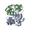
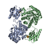
- PDB-1pxd: Crystal structure of the complex of jacalin with meso-tetrasulpho... -
+ Open data

- Basic information
| Entry | Database: PDB / ID: 1pxd | ||||||
|---|---|---|---|---|---|---|---|
| Title | Crystal structure of the complex of jacalin with meso-tetrasulphonatophenylporphyrin. | ||||||
 Components Components |
| ||||||
 Keywords Keywords | SUGAR BINDING PROTEIN / lectin / porphyrin | ||||||
| Function / homology |  Function and homology information Function and homology information | ||||||
| Biological species |   Artocarpus integer (chempedak) Artocarpus integer (chempedak) | ||||||
| Method |  X-RAY DIFFRACTION / MOLECULAR REPLACEMENT / Resolution: 1.8 Å X-RAY DIFFRACTION / MOLECULAR REPLACEMENT / Resolution: 1.8 Å | ||||||
 Authors Authors | Goel, M. / Anuradha, P. / Kaur, K.J. / Maiya, B.G. / Swamy, M.J. / Salunke, D.M. | ||||||
 Citation Citation | Journal: Acta Crystallogr.,Sect.D / Year: 2004 Title: Porphyrin binding to jacalin is facilitated by the inherent plasticity of the carbohydrate-binding site: novel mode of lectin-ligand interaction. Authors: Goel, M. / Anuradha, P. / Kaur, K.J. / Maiya, B.G. / Swamy, M.J. / Salunke, D.M. #1: Journal: J.Biol.Chem. / Year: 2001Title: Functional equality in the absence of structural similarity: an added dimension to molecular mimicry Authors: Goel, M. / Jain, D. / Kaur, K.J. / Kenoth, R. / Maiya, B.G. / Swamy, M.J. / Salunke, D.M. | ||||||
| History |
|
- Structure visualization
| Structure viewer | Molecule: MolmilJmol/JSmol |
|---|
- Downloads & links
-Download
| PDBx/mmCIF format | 1pxd.cif.gz | 46 KB | Display | PDBx/mmCIF format |
|---|---|---|---|---|
| PDB format | pdb1pxd.ent.gz | 32.6 KB | Display | PDB format |
| PDBx/mmJSON format | 1pxd.json.gz | Tree view | PDBx/mmJSON format | |
| Others |  Other downloads Other downloads |
-Validation report
| Arichive directory | https://data.pdbj.org/pub/pdb/validation_reports/px/1pxdftp://data.pdbj.org/pub/pdb/validation_reports/px/1pxd | HTTPS FTP |
|---|
-Related structure data
| Related structure data | 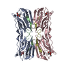 1jacS S: Starting model for refinement |
|---|---|
| Similar structure data |


-Links
 PDBj
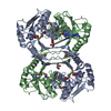
PDBj- Assembly
| Deposited unit |
| ||||||||
|---|---|---|---|---|---|---|---|---|---|
| 1 |
| ||||||||
| Unit cell |
|
-Components
| #1: Protein | Mass: 14673.479 Da / Num. of mol.: 1 / Source method: isolated from a natural source / Source: (natural) Artocarpus integer (chempedak) / References: UniProt: P18670 | ||
|---|---|---|---|
| #2: Protein/peptide | Mass: 2059.257 Da / Num. of mol.: 1 / Source method: isolated from a natural source / Source: (natural) Artocarpus integer (chempedak) / References: UniProt: P18673 | ||
| #3: Chemical | 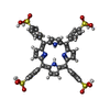  Mass: 939.020 Da / Num. of mol.: 2 / Source method: obtained synthetically / Formula: C44H34N4O12S4 Mass: 939.020 Da / Num. of mol.: 2 / Source method: obtained synthetically / Formula: C44H34N4O12S4#4: Water | ChemComp-HOH / |  Mass: 18.015 Da / Num. of mol.: 57 / Source method: isolated from a natural source / Formula: H2O Mass: 18.015 Da / Num. of mol.: 57 / Source method: isolated from a natural source / Formula: H2O |
-Experimental details
-Experiment
| Experiment | Method: X-RAY DIFFRACTION / Number of used crystals: 1 |
|---|
- Sample preparation
| Crystal | Density Matthews: 3.8 Å3/Da / Density % sol: 67.9 % |
|---|---|
| Crystal grow | Temperature: 298 K / Method: vapor diffusion, hanging drop / pH: 7.2 Details: Ammonium sulphate and Sodium chloride, pH 7.2, VAPOR DIFFUSION, HANGING DROP, temperature 298K |
-Data collection
| Diffraction | Mean temperature: 298 K |
|---|---|
| Diffraction source | Source: ROTATING ANODE / Type: RIGAKU RU200 / Wavelength: 1.5418 Å |
| Detector | Type: MARRESEARCH / Detector: IMAGE PLATE / Date: Feb 16, 2002 |
| Radiation | Monochromator: Graphite / Protocol: SINGLE WAVELENGTH / Monochromatic (M) / Laue (L): M / Scattering type: x-ray |
| Radiation wavelength | Wavelength: 1.5418 Å / Relative weight: 1 |
| Reflection | Resolution: 1.8→100 Å / Num. all: 23948 / Num. obs: 23948 / % possible obs: 87.9 % / Observed criterion σ(F): 0 / Observed criterion σ(I): 0 / Redundancy: 4.8 % / Rmerge(I) obs: 0.061 |
| Reflection shell | Resolution: 1.8→1.86 Å / Redundancy: 4.8 % / Rmerge(I) obs: 0.613 / Mean I/σ(I) obs: 10.8 / Num. unique all: 23948 / % possible all: 79.9 |
- Processing
| Software |
| |||||||||||||||||||||||||
|---|---|---|---|---|---|---|---|---|---|---|---|---|---|---|---|---|---|---|---|---|---|---|---|---|---|---|
| Refinement | Method to determine structure: MOLECULAR REPLACEMENT Starting model: 1JAC Resolution: 1.8→100 Å / σ(F): 0
| |||||||||||||||||||||||||
| Refinement step | Cycle: LAST / Resolution: 1.8→100 Å
|
