 Movie
Movie Controller


+ Open data

- Basic information
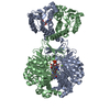
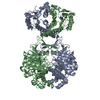
| Entry | Database: PDB / ID: 1pkx | |||||||||
|---|---|---|---|---|---|---|---|---|---|---|
| Title | Crystal Structure of human ATIC in complex with XMP | |||||||||
 Components Components | Bifunctional purine biosynthesis protein PURH | |||||||||
 Keywords Keywords | TRANSFERASE / HYDROLASE / ATIC / AICAR transformylase / IMP Cyclohydrolase / xanthosine monophosphate / purine biosynthesis | |||||||||
| Function / homology |  Function and homology information Function and homology informationphosphoribosylaminoimidazolecarboxamide formyltransferase / phosphoribosylaminoimidazolecarboxamide formyltransferase activity / IMP cyclohydrolase / IMP cyclohydrolase activity / brainstem development / 'de novo' XMP biosynthetic process / Purine ribonucleoside monophosphate biosynthesis / 'de novo' AMP biosynthetic process / GMP biosynthetic process / cellular response to interleukin-7 ...phosphoribosylaminoimidazolecarboxamide formyltransferase / phosphoribosylaminoimidazolecarboxamide formyltransferase activity / IMP cyclohydrolase / IMP cyclohydrolase activity / brainstem development / 'de novo' XMP biosynthetic process / Purine ribonucleoside monophosphate biosynthesis / 'de novo' AMP biosynthetic process / GMP biosynthetic process / cellular response to interleukin-7 / 'de novo' IMP biosynthetic process / nucleobase-containing compound metabolic process / dihydrofolate metabolic process / animal organ regeneration / tetrahydrofolate biosynthetic process / cerebellum development / cerebral cortex development / Signaling by ALK fusions and activated point mutants / cadherin binding / protein homodimerization activity / extracellular exosome / membrane / plasma membrane / cytosol Similarity search - Function | |||||||||
| Biological species |  Homo sapiens (human) Homo sapiens (human) | |||||||||
| Method |  X-RAY DIFFRACTION / SYNCHROTRON / MOLECULAR REPLACEMENT / Resolution: 1.9 Å X-RAY DIFFRACTION / SYNCHROTRON / MOLECULAR REPLACEMENT / Resolution: 1.9 Å | |||||||||
 Authors Authors | Wolan, D.W. / Cheong, C.G. / Greasley, S.E. / Wilson, I.A. | |||||||||
 Citation Citation | Journal: Biochemistry / Year: 2004 Title: Structural Insights into the Human and Avian IMP Cyclohydrolase Mechanism via Crystal Structures with the Bound XMP Inhibitor. Authors: Wolan, D.W. / Cheong, C.G. / Greasley, S.E. / Wilson, I.A. | |||||||||
| History |
|
- Structure visualization
| Structure viewer | Molecule: MolmilJmol/JSmol |
|---|
- Downloads & links
-Download
| PDBx/mmCIF format | 1pkx.cif.gz | 465.5 KB | Display | PDBx/mmCIF format |
|---|---|---|---|---|
| PDB format | pdb1pkx.ent.gz | 376.9 KB | Display | PDB format |
| PDBx/mmJSON format | 1pkx.json.gz | Tree view | PDBx/mmJSON format | |
| Others |  Other downloads Other downloads |
-Validation report
| Arichive directory | https://data.pdbj.org/pub/pdb/validation_reports/pk/1pkxftp://data.pdbj.org/pub/pdb/validation_reports/pk/1pkx | HTTPS FTP |
|---|
-Related structure data
| Related structure data | 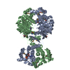 1g8mS S: Starting model for refinement |
|---|---|
| Similar structure data |


-Links
 PDBj
PDBj
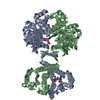
- Assembly
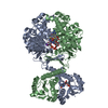
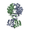
| Deposited unit | 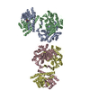
| ||||||||
|---|---|---|---|---|---|---|---|---|---|
| 1 | 
| ||||||||
| 2 | 
| ||||||||
| Unit cell |
| ||||||||
| Details | biological unit is a homodimer and AU contains two dimers |
-Components
| #1: Protein | Mass: 64693.719 Da / Num. of mol.: 4 Source method: isolated from a genetically manipulated source Details: Includes: Phosphoribosylaminoimidazolecarboxamide formyltransferase (AICAR transformylase) and IMP cyclohydrolase (Inosinicase) (IMP synthetase) Source: (gene. exp.) Homo sapiens (human) / Gene: ATIC / Production host:  References: UniProt: P31939, phosphoribosylaminoimidazolecarboxamide formyltransferase, IMP cyclohydrolase #2: Chemical | ChemComp-K /   Mass: 39.098 Da / Num. of mol.: 4 / Source method: obtained synthetically / Formula: K Mass: 39.098 Da / Num. of mol.: 4 / Source method: obtained synthetically / Formula: K#3: Chemical | 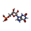  Mass: 365.213 Da / Num. of mol.: 2 / Source method: obtained synthetically / Formula: C10H14N4O9P Mass: 365.213 Da / Num. of mol.: 2 / Source method: obtained synthetically / Formula: C10H14N4O9P#4: Water | ChemComp-HOH / |  Mass: 18.015 Da / Num. of mol.: 977 / Source method: isolated from a natural source / Formula: H2O Mass: 18.015 Da / Num. of mol.: 977 / Source method: isolated from a natural source / Formula: H2OHas protein modification | N | |
|---|
-Experimental details
-Experiment
| Experiment | Method: X-RAY DIFFRACTION / Number of used crystals: 1 |
|---|
- Sample preparation
| Crystal | Density Matthews: 2.52 Å3/Da / Density % sol: 51.18 % | |||||||||||||||||||||||||||||||||||||||||||||||||
|---|---|---|---|---|---|---|---|---|---|---|---|---|---|---|---|---|---|---|---|---|---|---|---|---|---|---|---|---|---|---|---|---|---|---|---|---|---|---|---|---|---|---|---|---|---|---|---|---|---|---|
| Crystal grow | Temperature: 295 K / Method: vapor diffusion, sitting drop Details: Polyethylene glycol 3000, pH 7.5-8.0, VAPOR DIFFUSION, SITTING DROP, temperature 295K | |||||||||||||||||||||||||||||||||||||||||||||||||
| Crystal grow | *PLUS Temperature: 22 ℃ / Method: vapor diffusion, sitting drop / PH range low: 8 / PH range high: 7.5 | |||||||||||||||||||||||||||||||||||||||||||||||||
| Components of the solutions | *PLUS
|
-Data collection
| Diffraction | Mean temperature: 100 K |
|---|---|
| Diffraction source | Source: SYNCHROTRON / Site: SSRL  / Beamline: BL9-2 / Beamline: BL9-2 |
| Radiation | Protocol: SINGLE WAVELENGTH / Monochromatic (M) / Laue (L): M / Scattering type: x-ray |
| Radiation wavelength | Relative weight: 1 |
| Reflection | Resolution: 1.9→50 Å / Num. obs: 177135 / % possible obs: 87.7 % / Redundancy: 2.1 % / Rsym value: 0.053 / Net I/σ(I): 15 |
| Reflection shell | Resolution: 1.9→1.97 Å / Redundancy: 1.8 % / Mean I/σ(I) obs: 2.1 / Rsym value: 0.394 / % possible all: 47.8 |
| Reflection | *PLUS Rmerge(I) obs: 0.053 |
| Reflection shell | *PLUS % possible obs: 47.8 % / Num. unique obs: 9598 / Rmerge(I) obs: 0.394 |
- Processing
| Software |
| ||||||||||||||||
|---|---|---|---|---|---|---|---|---|---|---|---|---|---|---|---|---|---|
| Refinement | Method to determine structure: MOLECULAR REPLACEMENT Starting model: PDB entry 1G8M Resolution: 1.9→50 Å / σ(F): 0 / Stereochemistry target values: Engh & Huber
| ||||||||||||||||
| Refinement step | Cycle: LAST / Resolution: 1.9→50 Å
| ||||||||||||||||
| Refine LS restraints |
| ||||||||||||||||
| Refinement | *PLUS Lowest resolution: 50 Å / % reflection Rfree: 5.2 % | ||||||||||||||||
| Solvent computation | *PLUS | ||||||||||||||||
| Displacement parameters | *PLUS |
