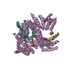
 Movie
Movie Controller


+ Open data

- Basic information
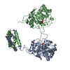
| Entry | Database: PDB / ID: 1pgr | ||||||
|---|---|---|---|---|---|---|---|
| Title | 2:2 COMPLEX OF G-CSF WITH ITS RECEPTOR | ||||||
 Components Components |
| ||||||
 Keywords Keywords | CYTOKINE / CLASS1 CYTOKINE / HEMATOPOIETIC RECEPTOR / SIGNAL TRANSDUCTION | ||||||
| Function / homology |  Function and homology information Function and homology informationgranulocyte colony-stimulating factor binding / granulocyte colony-stimulating factor receptor binding / regulation of myeloid cell differentiation / granulocyte colony-stimulating factor signaling pathway / positive regulation of myeloid cell differentiation / granulocyte differentiation / amelogenesis / regulation of actin filament organization / cytokine receptor activity / Other interleukin signaling ...granulocyte colony-stimulating factor binding / granulocyte colony-stimulating factor receptor binding / regulation of myeloid cell differentiation / granulocyte colony-stimulating factor signaling pathway / positive regulation of myeloid cell differentiation / granulocyte differentiation / amelogenesis / regulation of actin filament organization / cytokine receptor activity / Other interleukin signaling / cellular response to cytokine stimulus / Interleukin-10 signaling / positive regulation of actin filament polymerization / Signaling by CSF3 (G-CSF) / neutrophil chemotaxis / endocytic vesicle lumen / lysosomal lumen / cytokine activity / growth factor activity / Inactivation of CSF3 (G-CSF) signaling / cytokine-mediated signaling pathway / endocytic vesicle membrane / cellular response to lipopolysaccharide / response to ethanol / positive regulation of phosphatidylinositol 3-kinase/protein kinase B signal transduction / cell adhesion / immune response / external side of plasma membrane / positive regulation of cell population proliferation / enzyme binding / positive regulation of transcription by RNA polymerase II / : / extracellular region / plasma membrane Similarity search - Function | ||||||
| Biological species |  Homo sapiens (human) Homo sapiens (human) | ||||||
| Method |  X-RAY DIFFRACTION / SYNCHROTRON / MOLECULAR REPLACEMENT / Resolution: 3.5 Å X-RAY DIFFRACTION / SYNCHROTRON / MOLECULAR REPLACEMENT / Resolution: 3.5 Å | ||||||
 Authors Authors | Aritomi, M. / Kunishima, N. / Okamoto, T. / Kuroki, R. / Ota, Y. / Morikawa, K. | ||||||
 Citation Citation | Journal: Nature / Year: 1999 Title: Atomic structure of the GCSF-receptor complex showing a new cytokine-receptor recognition scheme. Authors: Aritomi, M. / Kunishima, N. / Okamoto, T. / Kuroki, R. / Ota, Y. / Morikawa, K. | ||||||
| History |
|
- Structure visualization
| Structure viewer | Molecule: MolmilJmol/JSmol |
|---|
- Downloads & links
-Download
| PDBx/mmCIF format | 1pgr.cif.gz | 282.8 KB | Display | PDBx/mmCIF format |
|---|---|---|---|---|
| PDB format | pdb1pgr.ent.gz | 234 KB | Display | PDB format |
| PDBx/mmJSON format | 1pgr.json.gz | Tree view | PDBx/mmJSON format | |
| Others |  Other downloads Other downloads |
-Validation report
| Arichive directory | https://data.pdbj.org/pub/pdb/validation_reports/pg/1pgrftp://data.pdbj.org/pub/pdb/validation_reports/pg/1pgr | HTTPS FTP |
|---|
-Related structure data
| Related structure data |  1cd9C 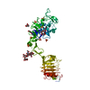 1igrS S: Starting model for refinement C: citing same article ( |
|---|---|
| Similar structure data |


-Links
 PDBj
PDBj


- Assembly
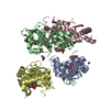
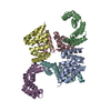
| Deposited unit | 
| ||||||||||||||||||||
|---|---|---|---|---|---|---|---|---|---|---|---|---|---|---|---|---|---|---|---|---|---|
| 1 | 
| ||||||||||||||||||||
| 2 | 
| ||||||||||||||||||||
| Unit cell |
| ||||||||||||||||||||
| Noncrystallographic symmetry (NCS) | NCS oper:
|
-Components
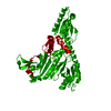
| #1: Protein | Mass: 18816.760 Da / Num. of mol.: 4 Source method: isolated from a genetically manipulated source Source: (gene. exp.) Homo sapiens (human) / Cellular location: EXTRACELLULAR / Production host:  #2: Protein | Mass: 24460.264 Da / Num. of mol.: 4 / Fragment: CRH REGION (BN DOMAIN:H1-108, BC DOMAIN:H109-215) Source method: isolated from a genetically manipulated source Source: (gene. exp.)   Spodoptera frugiperda (fall armyworm) / References: UniProt: P40223 Spodoptera frugiperda (fall armyworm) / References: UniProt: P40223Has protein modification | Y | |
|---|
-Experimental details
-Experiment
| Experiment | Method: X-RAY DIFFRACTION / Number of used crystals: 1 |
|---|
- Sample preparation
| Crystal | Density Matthews: 4.1 Å3/Da / Density % sol: 70 % |
|---|---|
| Crystal grow | pH: 7.5 / Details: pH 7.5 |
| Crystal grow | *PLUS Method: unknown / Details: manuscript in preparation |
-Data collection
| Diffraction | Mean temperature: 100 K |
|---|---|
| Diffraction source | Source: SYNCHROTRON / Site: Photon Factory  / Beamline: BL-6B / Wavelength: 1 / Beamline: BL-6B / Wavelength: 1 |
| Detector | Detector: IMAGE PLATE / Date: Dec 1, 1997 / Details: BENT MIRROR |
| Radiation | Monochromator: SI(111) / Protocol: SINGLE WAVELENGTH / Monochromatic (M) / Laue (L): M / Scattering type: x-ray |
| Radiation wavelength | Wavelength: 1 Å / Relative weight: 1 |
| Reflection | Resolution: 3.5→50 Å / Num. obs: 26681 / % possible obs: 69.1 % / Redundancy: 6.5 % / Biso Wilson estimate: 61.5 Å2 / Rmerge(I) obs: 0.198 |
| Reflection | *PLUS Num. obs: 26690 / Num. measured all: 173168 |
- Processing
| Software |
| ||||||||||||||||||||||||||||||||||||||||||||||||||||||||||||
|---|---|---|---|---|---|---|---|---|---|---|---|---|---|---|---|---|---|---|---|---|---|---|---|---|---|---|---|---|---|---|---|---|---|---|---|---|---|---|---|---|---|---|---|---|---|---|---|---|---|---|---|---|---|---|---|---|---|---|---|---|---|
| Refinement | Method to determine structure: MOLECULAR REPLACEMENT Starting model: 1IGR Resolution: 3.5→8 Å / σ(F): 0 Details: RIGID-BODY REFINEMENT THE 107TH VALINES IN CHAINS B,D,F,H WERE REMOVED FOR THE RIGID-BODY REFINEMENT. INITIAL MODEL WAS OBTAINED BY A MOLECULAR REPLACEMENT OF 1IGR. A RIGID-BODY REFINEMENT ...Details: RIGID-BODY REFINEMENT THE 107TH VALINES IN CHAINS B,D,F,H WERE REMOVED FOR THE RIGID-BODY REFINEMENT. INITIAL MODEL WAS OBTAINED BY A MOLECULAR REPLACEMENT OF 1IGR. A RIGID-BODY REFINEMENT WAS APPLIED TO THE MODEL AS INDEPENDENT 12 GROUPS. FURTHER POSITIONAL OR B-FACTOR REFINEMENTS FOR INDIVIDUAL ATOMS WERE NOT APPLIED.
| ||||||||||||||||||||||||||||||||||||||||||||||||||||||||||||
| Displacement parameters | Biso mean: 36.9 Å2 | ||||||||||||||||||||||||||||||||||||||||||||||||||||||||||||
| Refinement step | Cycle: LAST / Resolution: 3.5→8 Å
| ||||||||||||||||||||||||||||||||||||||||||||||||||||||||||||
| Refine LS restraints |
| ||||||||||||||||||||||||||||||||||||||||||||||||||||||||||||
| LS refinement shell | Resolution: 3.5→3.64 Å / Total num. of bins used: 8
| ||||||||||||||||||||||||||||||||||||||||||||||||||||||||||||
| Xplor file | Serial no: 1 / Param file: PARHCSDX.PRO / Topol file: TOPHCSDX.PRO |
