 Movie
Movie Controller

[English] 日本語
 Yorodumi
- PDB-1p3t: Crystal Structures of the NO-and CO-Bound Heme Oxygenase From Nei... -
+ Open data

- Basic information
| Entry | Database: PDB / ID: 1p3t | ||||||
|---|---|---|---|---|---|---|---|
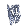
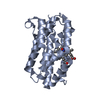
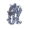
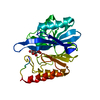
| Title | Crystal Structures of the NO-and CO-Bound Heme Oxygenase From Neisseria Meningitidis: Implications for Oxygen Activation | ||||||
 Components Components | Heme oxygenase 1 | ||||||
 Keywords Keywords | OXIDOREDUCTASE / heme oxygenase / heme degradation | ||||||
| Function / homology |  Function and homology information Function and homology informationheme oxidation / heme oxygenase (decyclizing) activity / metal ion binding Similarity search - Function | ||||||
| Biological species |  Neisseria meningitidis (bacteria) Neisseria meningitidis (bacteria) | ||||||
| Method |  X-RAY DIFFRACTION / MOLECULAR REPLACEMENT / Resolution: 2.1 Å X-RAY DIFFRACTION / MOLECULAR REPLACEMENT / Resolution: 2.1 Å | ||||||
 Authors Authors | Friedman, J. / Lad, L. / Deshmukh, R. / Li, H. / Wilks, A. / Poulos, T.L. | ||||||
 Citation Citation | Journal: J.Biol.Chem. / Year: 2003 Title: Crystal structures of the NO- and CO-bound heme oxygenase from Neisseriae meningitidis. Implications for O2 activation Authors: Friedman, J. / Lad, L. / Deshmukh, R. / Li, H. / Wilks, A. / Poulos, T.L. | ||||||
| History |
|
- Structure visualization
| Structure viewer | Molecule: MolmilJmol/JSmol |
|---|
- Downloads & links
-Download
| PDBx/mmCIF format | 1p3t.cif.gz | 54.7 KB | Display | PDBx/mmCIF format |
|---|---|---|---|---|
| PDB format | pdb1p3t.ent.gz | 38.7 KB | Display | PDB format |
| PDBx/mmJSON format | 1p3t.json.gz | Tree view | PDBx/mmJSON format | |
| Others |  Other downloads Other downloads |
-Validation report
| Arichive directory | https://data.pdbj.org/pub/pdb/validation_reports/p3/1p3tftp://data.pdbj.org/pub/pdb/validation_reports/p3/1p3t | HTTPS FTP |
|---|
-Related structure data
| Related structure data | 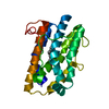 1p3uC  1p3vC 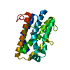 1j77S C: citing same article ( S: Starting model for refinement |
|---|---|
| Similar structure data |


-Links
 PDBj
PDBj- Assembly
| Deposited unit | 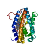
| ||||||||||
|---|---|---|---|---|---|---|---|---|---|---|---|
| 1 |
| ||||||||||
| Unit cell |
|
-Components
| #1: Protein | Mass: 23609.611 Da / Num. of mol.: 1 Source method: isolated from a genetically manipulated source Details: Heme-complexed / Source: (gene. exp.) Neisseria meningitidis (bacteria) / Gene: hemO / Plasmid: pWMZ1651 / Species (production host): Escherichia coli / Production host: References: UniProt: Q9RGD9, heme oxygenase (biliverdin-producing) |
|---|---|
| #2: Chemical | ChemComp-HEM /   Mass: 616.487 Da / Num. of mol.: 1 / Source method: obtained synthetically / Formula: C34H32FeN4O4 Mass: 616.487 Da / Num. of mol.: 1 / Source method: obtained synthetically / Formula: C34H32FeN4O4 |
| #3: Water | ChemComp-HOH /  Mass: 18.015 Da / Num. of mol.: 62 / Source method: isolated from a natural source / Formula: H2O Mass: 18.015 Da / Num. of mol.: 62 / Source method: isolated from a natural source / Formula: H2O |
-Experimental details
-Experiment
| Experiment | Method: X-RAY DIFFRACTION / Number of used crystals: 1 |
|---|
- Sample preparation
| Crystal | Density Matthews: 2.16 Å3/Da / Density % sol: 43.12 % | ||||||||||||||||||||||||||||||
|---|---|---|---|---|---|---|---|---|---|---|---|---|---|---|---|---|---|---|---|---|---|---|---|---|---|---|---|---|---|---|---|
| Crystal grow | Temperature: 298 K / Method: vapor diffusion, hanging drop / pH: 8.5 Details: tris-HCl, sodium acetate, PEG 3350, pH 8.5, VAPOR DIFFUSION, HANGING DROP, temperature 298K | ||||||||||||||||||||||||||||||
| Crystal grow | *PLUS Method: vapor diffusion, sitting drop | ||||||||||||||||||||||||||||||
| Components of the solutions | *PLUS
|
-Data collection
| Diffraction | Mean temperature: 119 K |
|---|---|
| Diffraction source | Source: ROTATING ANODE / Type: RIGAKU / Wavelength: 1.5418 Å |
| Detector | Type: RIGAKU RAXIS IV / Detector: IMAGE PLATE / Date: Jul 14, 2002 / Details: mirrors |
| Radiation | Monochromator: yale mirrors / Protocol: SINGLE WAVELENGTH / Monochromatic (M) / Laue (L): M / Scattering type: x-ray |
| Radiation wavelength | Wavelength: 1.5418 Å / Relative weight: 1 |
| Reflection | Resolution: 2.1→50 Å / Num. obs: 12602 / % possible obs: 94.6 % / Observed criterion σ(I): 2 / Redundancy: 9.5 % / Rmerge(I) obs: 0.051 / Rsym value: 0.067 / Net I/σ(I): 38.5 |
| Reflection shell | Resolution: 2.1→2.18 Å / Redundancy: 4.67 % / Rmerge(I) obs: 0.611 / Mean I/σ(I) obs: 2.37 / Num. unique all: 1284 / Rsym value: 0.555 / % possible all: 97.8 |
| Reflection | *PLUS Num. obs: 31166 / % possible obs: 96.7 % / Num. measured all: 226194 |
| Reflection shell | *PLUS % possible obs: 97.8 % |
- Processing
| Software |
| |||||||||||||||||||||||||
|---|---|---|---|---|---|---|---|---|---|---|---|---|---|---|---|---|---|---|---|---|---|---|---|---|---|---|
| Refinement | Method to determine structure: MOLECULAR REPLACEMENT Starting model: 1J77 Resolution: 2.1→50 Å / Isotropic thermal model: isotropic / Cross valid method: THROUGHOUT / σ(F): 0 / Stereochemistry target values: Engh & Huber
| |||||||||||||||||||||||||
| Displacement parameters | Biso mean: 46.44 Å2 | |||||||||||||||||||||||||
| Refinement step | Cycle: LAST / Resolution: 2.1→50 Å
| |||||||||||||||||||||||||
| Refine LS restraints |
| |||||||||||||||||||||||||
| Refinement | *PLUS % reflection Rfree: 5 % | |||||||||||||||||||||||||
| Solvent computation | *PLUS | |||||||||||||||||||||||||
| Displacement parameters | *PLUS | |||||||||||||||||||||||||
| Refine LS restraints | *PLUS Type: c_angle_deg / Dev ideal: 1.374 |
