 Movie
Movie Controller

[English] 日本語
 Yorodumi
- PDB-1j77: Crystal Structure of Gram-negative Bacterial Heme Oxygenase Compl... -
+ Open data

- Basic information
| Entry | Database: PDB / ID: 1j77 | ||||||
|---|---|---|---|---|---|---|---|
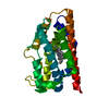
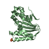
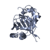
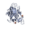
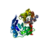
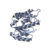
| Title | Crystal Structure of Gram-negative Bacterial Heme Oxygenase Complexed with Heme | ||||||
 Components Components | HemO | ||||||
 Keywords Keywords | OXIDOREDUCTASE / proximal histidine / distal helix | ||||||
| Function / homology |  Function and homology information Function and homology informationheme oxidation / heme oxygenase (decyclizing) activity / metal ion binding Similarity search - Function | ||||||
| Biological species |  Neisseria meningitidis (bacteria) Neisseria meningitidis (bacteria) | ||||||
| Method |  X-RAY DIFFRACTION / SYNCHROTRON / MAD / Resolution: 1.5 Å X-RAY DIFFRACTION / SYNCHROTRON / MAD / Resolution: 1.5 Å | ||||||
 Authors Authors | Schuller, D.J. / Zhu, W. / Stojiljkovic, I. / Wilks, A. / Poulos, T.L. | ||||||
 Citation Citation | Journal: Biochemistry / Year: 2001 Title: Crystal structure of heme oxygenase from the gram-negative pathogen Neisseria meningitidis and a comparison with mammalian heme oxygenase-1. Authors: Schuller, D.J. / Zhu, W. / Stojiljkovic, I. / Wilks, A. / Poulos, T.L. | ||||||
| History |
|
- Structure visualization
| Structure viewer | Molecule: MolmilJmol/JSmol |
|---|
- Downloads & links
-Download
| PDBx/mmCIF format | 1j77.cif.gz | 102.9 KB | Display | PDBx/mmCIF format |
|---|---|---|---|---|
| PDB format | pdb1j77.ent.gz | 79.3 KB | Display | PDB format |
| PDBx/mmJSON format | 1j77.json.gz | Tree view | PDBx/mmJSON format | |
| Others |  Other downloads Other downloads |
-Validation report
| Arichive directory | https://data.pdbj.org/pub/pdb/validation_reports/j7/1j77ftp://data.pdbj.org/pub/pdb/validation_reports/j7/1j77 | HTTPS FTP |
|---|
-Related structure data
| Related structure data | |
|---|---|
| Similar structure data |


-Links
 PDBj
PDBj- Assembly
| Deposited unit | 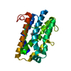
| ||||||||
|---|---|---|---|---|---|---|---|---|---|
| 1 |
| ||||||||
| Unit cell |
| ||||||||
| Components on special symmetry positions |
| ||||||||
| Details | the enzyme is a monomer |
-Components
| #1: Protein | Mass: 23609.611 Da / Num. of mol.: 1 Source method: isolated from a genetically manipulated source Source: (gene. exp.) Neisseria meningitidis (bacteria) / Gene: hemO / Plasmid: pWMZ1651 / Species (production host): Escherichia coli / Production host: References: UniProt: Q9RGD9, heme oxygenase (biliverdin-producing) |
|---|---|
| #2: Chemical | ChemComp-HEM /   Mass: 616.487 Da / Num. of mol.: 1 / Source method: obtained synthetically / Formula: C34H32FeN4O4 Mass: 616.487 Da / Num. of mol.: 1 / Source method: obtained synthetically / Formula: C34H32FeN4O4 |
| #3: Water | ChemComp-HOH /  Mass: 18.015 Da / Num. of mol.: 244 / Source method: isolated from a natural source / Formula: H2O Mass: 18.015 Da / Num. of mol.: 244 / Source method: isolated from a natural source / Formula: H2O |
-Experimental details
-Experiment
| Experiment | Method: X-RAY DIFFRACTION / Number of used crystals: 1 |
|---|
- Sample preparation
| Crystal | Density Matthews: 2.12 Å3/Da / Density % sol: 43 % | |||||||||||||||||||||||||
|---|---|---|---|---|---|---|---|---|---|---|---|---|---|---|---|---|---|---|---|---|---|---|---|---|---|---|
| Crystal grow | Temperature: 298 K / Method: vapor diffusion, sitting drop / pH: 8.5 Details: PEG 3350, sodium acetate, Tris hydrochloride, pH 8.50, VAPOR DIFFUSION, SITTING DROP, temperature 298.0K | |||||||||||||||||||||||||
| Crystal grow | *PLUS pH: 8.5 | |||||||||||||||||||||||||
| Components of the solutions | *PLUS
|
-Data collection
| Diffraction | Mean temperature: 100 K | |||||||||||||||
|---|---|---|---|---|---|---|---|---|---|---|---|---|---|---|---|---|
| Diffraction source | Source: SYNCHROTRON / Site: SSRL  / Beamline: BL9-1 / Wavelength: 1.5498, 1.7377, 1.7401, 1.9075 / Beamline: BL9-1 / Wavelength: 1.5498, 1.7377, 1.7401, 1.9075 | |||||||||||||||
| Detector | Type: MARRESEARCH / Detector: IMAGE PLATE / Date: May 26, 2000 Details: Flat mirror(vertical) single crystal (Si311) bent monochromator (horizontal) | |||||||||||||||
| Radiation | Monochromator: bent crystal Si(311) / Protocol: SINGLE WAVELENGTH / Monochromatic (M) / Laue (L): M / Scattering type: x-ray | |||||||||||||||
| Radiation wavelength |
| |||||||||||||||
| Reflection | Resolution: 1.5→44.67 Å / Num. all: 31486 / Num. obs: 31486 / % possible obs: 0.946 % / Observed criterion σ(F): 0 / Observed criterion σ(I): 0 / Redundancy: 5.1 % / Biso Wilson estimate: 23.743 Å2 / Rmerge(I) obs: 0.047 / Net I/σ(I): 13.3 | |||||||||||||||
| Reflection shell | Resolution: 1.5→1.53 Å / Redundancy: 2.5 % / Rmerge(I) obs: 0.532 / Mean I/σ(I) obs: 2.1 / Num. unique all: 1381 / % possible all: 92.1 | |||||||||||||||
| Reflection | *PLUS % possible obs: 94.9 % / Num. measured all: 161249 | |||||||||||||||
| Reflection shell | *PLUS % possible obs: 92.1 % |
- Processing
| Software |
| |||||||||||||||||||||||||
|---|---|---|---|---|---|---|---|---|---|---|---|---|---|---|---|---|---|---|---|---|---|---|---|---|---|---|
| Refinement | Method to determine structure: MAD / Resolution: 1.5→53.45 Å / Isotropic thermal model: anisotropic / Cross valid method: THROUGHOUT / σ(F): 0 / σ(I): 0 / Stereochemistry target values: REFMAC5 / Details: Maximum likelihood based on F
| |||||||||||||||||||||||||
| Displacement parameters | Biso mean: 22.9 Å2
| |||||||||||||||||||||||||
| Refine analyze |
| |||||||||||||||||||||||||
| Refinement step | Cycle: LAST / Resolution: 1.5→53.45 Å
| |||||||||||||||||||||||||
| Refine LS restraints |
| |||||||||||||||||||||||||
| LS refinement shell | Resolution: 1.5→1.539 Å / Rfactor Rfree error: -0
| |||||||||||||||||||||||||
| Software | *PLUS Name: REFMAC / Version: 5 / Classification: refinement | |||||||||||||||||||||||||
| Refinement | *PLUS σ(F): 0 / % reflection Rfree: 5.1 % / Rfactor Rfree: 0.26 | |||||||||||||||||||||||||
| Solvent computation | *PLUS | |||||||||||||||||||||||||
| Displacement parameters | *PLUS Biso mean: 22.9 Å2 | |||||||||||||||||||||||||
| Refine LS restraints | *PLUS
|
