| 登録情報 | データベース: PDB / ID: 1p14
|
|---|
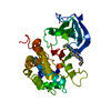
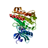
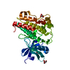
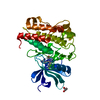
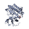
| タイトル | Crystal structure of a catalytic-loop mutant of the insulin receptor tyrosine kinase |
|---|
 要素 要素 | insulin receptor |
|---|
 キーワード キーワード | TRANSFERASE / RECEPTOR / TYROSINE KINASE / CATALYSIS / MUTANT |
|---|
| 機能・相同性 |  機能・相同性情報 機能・相同性情報
regulation of female gonad development / positive regulation of meiotic cell cycle / insulin-like growth factor II binding / positive regulation of developmental growth / male sex determination / insulin receptor complex / positive regulation of protein-containing complex disassembly / insulin-like growth factor I binding / insulin receptor activity / exocrine pancreas development ...regulation of female gonad development / positive regulation of meiotic cell cycle / insulin-like growth factor II binding / positive regulation of developmental growth / male sex determination / insulin receptor complex / positive regulation of protein-containing complex disassembly / insulin-like growth factor I binding / insulin receptor activity / exocrine pancreas development / dendritic spine maintenance / cargo receptor activity / insulin binding / adrenal gland development / PTB domain binding / neuronal cell body membrane / Signaling by Insulin receptor / IRS activation / positive regulation of respiratory burst / amyloid-beta clearance / regulation of embryonic development / positive regulation of receptor internalization / insulin receptor substrate binding / epidermis development / positive regulation of glycogen biosynthetic process / protein kinase activator activity / Signal attenuation / heart morphogenesis / transport across blood-brain barrier / phosphatidylinositol 3-kinase binding / Insulin receptor recycling / insulin-like growth factor receptor binding / dendrite membrane / neuron projection maintenance / positive regulation of mitotic nuclear division / receptor-mediated endocytosis / Insulin receptor signalling cascade / positive regulation of glycolytic process / positive regulation of D-glucose import across plasma membrane / learning / receptor protein-tyrosine kinase / caveola / receptor internalization / cellular response to growth factor stimulus / male gonad development / memory / cellular response to insulin stimulus / positive regulation of nitric oxide biosynthetic process / insulin receptor signaling pathway / late endosome / glucose homeostasis / amyloid-beta binding / protein autophosphorylation / PI5P, PP2A and IER3 Regulate PI3K/AKT Signaling / protein tyrosine kinase activity / lysosome / positive regulation of phosphatidylinositol 3-kinase/protein kinase B signal transduction / positive regulation of canonical NF-kappaB signal transduction / receptor complex / positive regulation of MAPK cascade / endosome membrane / positive regulation of cell migration / G protein-coupled receptor signaling pathway / protein domain specific binding / axon / external side of plasma membrane / positive regulation of cell population proliferation / regulation of DNA-templated transcription / symbiont entry into host cell / positive regulation of DNA-templated transcription / GTP binding / protein-containing complex binding / extracellular exosome / ATP binding / identical protein binding / membrane / plasma membrane類似検索 - 分子機能 Insulin receptor, trans-membrane domain / Insulin receptor trans-membrane segment / Tyrosine-protein kinase, insulin-like receptor / Tyrosine-protein kinase, receptor class II, conserved site / Receptor tyrosine kinase class II signature. / Receptor L-domain / Furin-like cysteine-rich domain / Receptor L-domain superfamily / Furin-like cysteine rich region / Receptor L domain ...Insulin receptor, trans-membrane domain / Insulin receptor trans-membrane segment / Tyrosine-protein kinase, insulin-like receptor / Tyrosine-protein kinase, receptor class II, conserved site / Receptor tyrosine kinase class II signature. / Receptor L-domain / Furin-like cysteine-rich domain / Receptor L-domain superfamily / Furin-like cysteine rich region / Receptor L domain / Furin-like repeat / Furin-like repeats / Fibronectin type III domain / Growth factor receptor cysteine-rich domain superfamily / : / Fibronectin type 3 domain / Fibronectin type-III domain profile. / Fibronectin type III / Fibronectin type III superfamily / Tyrosine-protein kinase, catalytic domain / Tyrosine kinase, catalytic domain / Tyrosine protein kinases specific active-site signature. / Tyrosine-protein kinase, active site / Serine-threonine/tyrosine-protein kinase, catalytic domain / Protein tyrosine and serine/threonine kinase / Phosphorylase Kinase; domain 1 / Phosphorylase Kinase; domain 1 / Transferase(Phosphotransferase) domain 1 / Transferase(Phosphotransferase); domain 1 / Protein kinase, ATP binding site / Protein kinases ATP-binding region signature. / Immunoglobulin-like fold / Protein kinase domain profile. / Protein kinase domain / Protein kinase-like domain superfamily / 2-Layer Sandwich / Orthogonal Bundle / Mainly Alpha / Alpha Beta類似検索 - ドメイン・相同性 |
|---|
| 生物種 |  Homo sapiens (ヒト) Homo sapiens (ヒト) |
|---|
| 手法 |  X線回折 / シンクロトロン / 分子置換 / 解像度: 1.9 Å X線回折 / シンクロトロン / 分子置換 / 解像度: 1.9 Å |
|---|
 データ登録者 データ登録者 | Li, S. / Covino, N.D. / Stein, E.G. / Till, J.H. / Hubbard, S.R. |
|---|
 引用 引用 | ジャーナル: J.Biol.Chem. / 年: 2003
タイトル: Structural and biochemical evidence for an autoinhibitory role for tyrosine 984 in the juxtamembrane region of the insulin receptor
著者: Li, S. / Covino, N.D. / Stein, E.G. / Till, J.H. / Hubbard, S.R. |
|---|
| 履歴 | | 登録 | 2003年4月11日 | 登録サイト: RCSB / 処理サイト: RCSB |
|---|
| 改定 1.0 | 2003年7月22日 | Provider: repository / タイプ: Initial release |
|---|
| 改定 1.1 | 2008年4月29日 | Group: Version format compliance |
|---|
| 改定 1.2 | 2011年7月13日 | Group: Version format compliance |
|---|
| 改定 1.3 | 2021年10月27日 | Group: Database references / カテゴリ: database_2 / struct_ref_seq_dif
Item: _database_2.pdbx_DOI / _database_2.pdbx_database_accession / _struct_ref_seq_dif.details |
|---|
| 改定 1.4 | 2023年8月16日 | Group: Data collection / Refinement description
カテゴリ: chem_comp_atom / chem_comp_bond / pdbx_initial_refinement_model |
|---|
|
|---|
 ムービー
ムービー コントローラー


 データを開く
 基本情報
基本情報 構造の表示
構造の表示 ダウンロードとリンク
 その他のダウンロード


 PDBj
PDBj


 集合体
集合体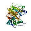

 Spodoptera frugiperda (ツマジロクサヨトウ)
 分子量: 18.015 Da / 分子数: 188 / 由来タイプ: 天然 / 式: H2O
 試料調製
試料調製 / ビームライン: X12C / 波長: 0.979 Å
 解析
解析