 Movie
Movie Controller

[English] 日本語
 Yorodumi
- PDB-1i44: CRYSTALLOGRAPHIC STUDIES OF AN ACTIVATION LOOP MUTANT OF THE INSU... -
+ Open data

- Basic information
| Entry | Database: PDB / ID: 1i44 | ||||||
|---|---|---|---|---|---|---|---|
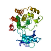
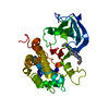
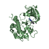
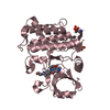
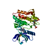
| Title | CRYSTALLOGRAPHIC STUDIES OF AN ACTIVATION LOOP MUTANT OF THE INSULIN RECEPTOR TYROSINE KINASE | ||||||
 Components Components | INSULIN RECEPTOR | ||||||
 Keywords Keywords | TRANSFERASE / protein tyrosine kinase / phosphotransferase | ||||||
| Function / homology |  Function and homology information Function and homology informationregulation of female gonad development / positive regulation of meiotic cell cycle / insulin-like growth factor II binding / positive regulation of developmental growth / insulin receptor complex / insulin-like growth factor I binding / insulin receptor activity / positive regulation of protein-containing complex disassembly / adrenal gland development / dendritic spine maintenance ...regulation of female gonad development / positive regulation of meiotic cell cycle / insulin-like growth factor II binding / positive regulation of developmental growth / insulin receptor complex / insulin-like growth factor I binding / insulin receptor activity / positive regulation of protein-containing complex disassembly / adrenal gland development / dendritic spine maintenance / insulin binding / cargo receptor activity / PTB domain binding / Signaling by Insulin receptor / IRS activation / neuronal cell body membrane / positive regulation of respiratory burst / amyloid-beta clearance / insulin receptor substrate binding / positive regulation of receptor internalization / heart morphogenesis / regulation of embryonic development / positive regulation of glycogen biosynthetic process / Signal attenuation / protein kinase activator activity / phosphatidylinositol 3-kinase binding / transport across blood-brain barrier / Insulin receptor recycling / insulin-like growth factor receptor binding / neuron projection maintenance / positive regulation of mitotic nuclear division / receptor-mediated endocytosis / positive regulation of glycolytic process / Insulin receptor signalling cascade / learning / dendrite membrane / positive regulation of D-glucose import across plasma membrane / male gonad development / receptor protein-tyrosine kinase / memory / caveola / cellular response to insulin stimulus / positive regulation of nitric oxide biosynthetic process / glucose homeostasis / insulin receptor signaling pathway / late endosome / protein autophosphorylation / amyloid-beta binding / PI5P, PP2A and IER3 Regulate PI3K/AKT Signaling / protein tyrosine kinase activity / positive regulation of MAPK cascade / positive regulation of phosphatidylinositol 3-kinase/protein kinase B signal transduction / positive regulation of canonical NF-kappaB signal transduction / signaling receptor complex / lysosome / endosome membrane / positive regulation of cell migration / G protein-coupled receptor signaling pathway / external side of plasma membrane / protein domain specific binding / axon / positive regulation of cell population proliferation / symbiont entry into host cell / regulation of DNA-templated transcription / positive regulation of DNA-templated transcription / GTP binding / protein-containing complex binding / extracellular exosome / ATP binding / membrane / identical protein binding / plasma membrane Similarity search - Function | ||||||
| Biological species |  Homo sapiens (human) Homo sapiens (human) | ||||||
| Method |  X-RAY DIFFRACTION / MOLECULAR REPLACEMENT / Resolution: 2.4 Å X-RAY DIFFRACTION / MOLECULAR REPLACEMENT / Resolution: 2.4 Å | ||||||
 Authors Authors | Till, J.H. / Ablooglu, A.J. / Frankel, M. / Kohanski, R.A. / Hubbard, S.R. | ||||||
 Citation Citation | Journal: J.Biol.Chem. / Year: 2001 Title: Crystallographic and solution studies of an activation loop mutant of the insulin receptor tyrosine kinase: insights into kinase mechanism. Authors: Till, J.H. / Ablooglu, A.J. / Frankel, M. / Bishop, S.M. / Kohanski, R.A. / Hubbard, S.R. | ||||||
| History |
|
- Structure visualization
| Structure viewer | Molecule: MolmilJmol/JSmol |
|---|
- Downloads & links
-Download
| PDBx/mmCIF format | 1i44.cif.gz | 75 KB | Display | PDBx/mmCIF format |
|---|---|---|---|---|
| PDB format | pdb1i44.ent.gz | 53 KB | Display | PDB format |
| PDBx/mmJSON format | 1i44.json.gz | Tree view | PDBx/mmJSON format | |
| Others |  Other downloads Other downloads |
-Validation report
| Arichive directory | https://data.pdbj.org/pub/pdb/validation_reports/i4/1i44ftp://data.pdbj.org/pub/pdb/validation_reports/i4/1i44 | HTTPS FTP |
|---|
-Related structure data
| Related structure data |  1irkS S: Starting model for refinement |
|---|---|
| Similar structure data |


-Links
 PDBj
PDBj


- Assembly
| Deposited unit | 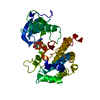
| ||||||||
|---|---|---|---|---|---|---|---|---|---|
| 1 |
| ||||||||
| Unit cell |
|
-Components
| #1: Protein | Mass: 34748.797 Da / Num. of mol.: 1 / Fragment: TYROSINE KINASE DOMAIN / Mutation: C981S, Y984PHE, D1161A Source method: isolated from a genetically manipulated source Source: (gene. exp.) Homo sapiens (human) / Production host:   Spodoptera frugiperda (fall armyworm) / References: UniProt: P06213, EC: 2.7.1.112 Spodoptera frugiperda (fall armyworm) / References: UniProt: P06213, EC: 2.7.1.112 |
|---|---|
| #2: Chemical | ChemComp-MG /   Mass: 24.305 Da / Num. of mol.: 1 / Source method: obtained synthetically / Formula: Mg Mass: 24.305 Da / Num. of mol.: 1 / Source method: obtained synthetically / Formula: Mg |
| #3: Chemical | ChemComp-ACP /   Mass: 505.208 Da / Num. of mol.: 1 / Source method: obtained synthetically / Formula: C11H18N5O12P3 / Comment: AMP-PCP, energy-carrying molecule analogue*YM Mass: 505.208 Da / Num. of mol.: 1 / Source method: obtained synthetically / Formula: C11H18N5O12P3 / Comment: AMP-PCP, energy-carrying molecule analogue*YM |
| #4: Water | ChemComp-HOH /  Mass: 18.015 Da / Num. of mol.: 141 / Source method: isolated from a natural source / Formula: H2O Mass: 18.015 Da / Num. of mol.: 141 / Source method: isolated from a natural source / Formula: H2O |
-Experimental details
-Experiment
| Experiment | Method: X-RAY DIFFRACTION / Number of used crystals: 1 |
|---|
- Sample preparation
| Crystal | Density Matthews: 2.58 Å3/Da / Density % sol: 52.4 % | ||||||||||||||||||||||||||||||||||||||||||||||||
|---|---|---|---|---|---|---|---|---|---|---|---|---|---|---|---|---|---|---|---|---|---|---|---|---|---|---|---|---|---|---|---|---|---|---|---|---|---|---|---|---|---|---|---|---|---|---|---|---|---|
| Crystal grow | Temperature: 293 K / Method: vapor diffusion, hanging drop / pH: 7.5 Details: PEG 8000, Tris-HCl, NaCl, dithiothreitol, pH 7.5, VAPOR DIFFUSION, HANGING DROP, temperature 293K | ||||||||||||||||||||||||||||||||||||||||||||||||
| Crystal | *PLUS Density % sol: 51 % | ||||||||||||||||||||||||||||||||||||||||||||||||
| Crystal grow | *PLUS Temperature: 20 ℃ | ||||||||||||||||||||||||||||||||||||||||||||||||
| Components of the solutions | *PLUS
|
-Data collection
| Diffraction | Mean temperature: 100 K |
|---|---|
| Diffraction source | Source: ROTATING ANODE / Type: RIGAKU RU200 / Wavelength: 1.5418 Å |
| Detector | Type: RIGAKU RAXIS II / Detector: IMAGE PLATE / Date: Jun 29, 2000 / Details: mirrors |
| Radiation | Monochromator: YALE MIRRORS / Protocol: SINGLE WAVELENGTH / Monochromatic (M) / Laue (L): M / Scattering type: x-ray |
| Radiation wavelength | Wavelength: 1.5418 Å / Relative weight: 1 |
| Reflection | Resolution: 2.4→30 Å / Num. all: 14350 / Num. obs: 14350 / % possible obs: 99 % / Observed criterion σ(F): 0 / Observed criterion σ(I): 0 / Redundancy: 3.2 % / Biso Wilson estimate: 17.5 Å2 / Rmerge(I) obs: 0.064 / Net I/σ(I): 11.6 |
| Reflection shell | Resolution: 2.4→2.49 Å / Rmerge(I) obs: 0.223 / % possible all: 93.9 |
| Reflection | *PLUS Lowest resolution: 30 Å / % possible obs: 99 % / Num. measured all: 76712 |
| Reflection shell | *PLUS % possible obs: 93.5 % |
- Processing
| Software |
| ||||||||||||||||||||||||||||||||||||||||||||||||||||||||||||||||||||||||||||||||
|---|---|---|---|---|---|---|---|---|---|---|---|---|---|---|---|---|---|---|---|---|---|---|---|---|---|---|---|---|---|---|---|---|---|---|---|---|---|---|---|---|---|---|---|---|---|---|---|---|---|---|---|---|---|---|---|---|---|---|---|---|---|---|---|---|---|---|---|---|---|---|---|---|---|---|---|---|---|---|---|---|---|
| Refinement | Method to determine structure: MOLECULAR REPLACEMENT Starting model: 1irk Resolution: 2.4→30 Å / Rfactor Rfree error: 0.01 / Data cutoff high absF: 362135.97 / Data cutoff low absF: 0 / Isotropic thermal model: RESTRAINED / Cross valid method: THROUGHOUT / σ(F): 0 / σ(I): 0 / Stereochemistry target values: ENGH AND HUBER, 1991
| ||||||||||||||||||||||||||||||||||||||||||||||||||||||||||||||||||||||||||||||||
| Solvent computation | Solvent model: FLAT MODEL / Bsol: 22.8 Å2 / ksol: 0.321 e/Å3 | ||||||||||||||||||||||||||||||||||||||||||||||||||||||||||||||||||||||||||||||||
| Displacement parameters | Biso mean: 25.9 Å2
| ||||||||||||||||||||||||||||||||||||||||||||||||||||||||||||||||||||||||||||||||
| Refine analyze |
| ||||||||||||||||||||||||||||||||||||||||||||||||||||||||||||||||||||||||||||||||
| Refinement step | Cycle: LAST / Resolution: 2.4→30 Å
| ||||||||||||||||||||||||||||||||||||||||||||||||||||||||||||||||||||||||||||||||
| Refine LS restraints |
| ||||||||||||||||||||||||||||||||||||||||||||||||||||||||||||||||||||||||||||||||
| LS refinement shell | Resolution: 2.4→2.55 Å / Rfactor Rfree error: 0.034 / Total num. of bins used: 6
| ||||||||||||||||||||||||||||||||||||||||||||||||||||||||||||||||||||||||||||||||
| Xplor file |
| ||||||||||||||||||||||||||||||||||||||||||||||||||||||||||||||||||||||||||||||||
| Software | *PLUS Name: CNS / Version: 0.5 / Classification: refinement | ||||||||||||||||||||||||||||||||||||||||||||||||||||||||||||||||||||||||||||||||
| Refinement | *PLUS Lowest resolution: 30 Å / σ(F): 0 / % reflection Rfree: 5 % / Rfactor obs: 0.21 / Rfactor Rwork: 0.21 | ||||||||||||||||||||||||||||||||||||||||||||||||||||||||||||||||||||||||||||||||
| Solvent computation | *PLUS | ||||||||||||||||||||||||||||||||||||||||||||||||||||||||||||||||||||||||||||||||
| Displacement parameters | *PLUS Biso mean: 25.9 Å2 | ||||||||||||||||||||||||||||||||||||||||||||||||||||||||||||||||||||||||||||||||
| Refine LS restraints | *PLUS
| ||||||||||||||||||||||||||||||||||||||||||||||||||||||||||||||||||||||||||||||||
| LS refinement shell | *PLUS Rfactor Rfree: 0.336 / % reflection Rfree: 4.8 % / Rfactor Rwork: 0.238 |
