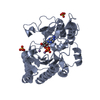
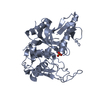
Entry Database : PDB / ID : 4ibmTitle Crystal structure of insulin receptor kinase domain in complex with an inhibitor Irfin-1 Insulin receptor Keywords / / / / / / Function / homology Function Domain/homology Component
/ / / / / / / / / / / / / / / / / / / / / / / / / / / / / / / / / / / / / / / / / / / / / / / / / / / / / / / / / / / / / / / / / / / / / / / / / / / / / / / / / / / / / / / / / / / / / / / / / / / / / / / / / / / / / / / / / / / / / / / / / / / / / / Biological species Homo sapiens (human)Method / / / Resolution : 1.8 Å Authors Wu, J. / Anastassiadis, T. / Duong-Ly, K.C. / Peterson, J.R. Journal : J.Biol.Chem. / Year : 2013Title : A highly selective dual insulin receptor (IR)/insulin-like growth factor 1 receptor (IGF-1R) inhibitor derived from an extracellular signal-regulated kinase (ERK) inhibitor.Authors : Anastassiadis, T. / Duong-Ly, K.C. / Deacon, S.W. / Lafontant, A. / Ma, H. / Devarajan, K. / Dunbrack, R.L. / Wu, J. / Peterson, J.R. History Deposition Dec 8, 2012 Deposition site / Processing site Revision 1.0 Aug 21, 2013 Provider / Type Revision 1.1 Aug 28, 2013 Group Revision 1.2 Nov 20, 2013 Group Revision 1.3 Nov 15, 2017 Group / Category / Item Revision 1.4 Feb 28, 2024 Group / Database references / Derived calculationsCategory chem_comp_atom / chem_comp_bond ... chem_comp_atom / chem_comp_bond / database_2 / struct_ref_seq_dif / struct_site Item _database_2.pdbx_DOI / _database_2.pdbx_database_accession ... _database_2.pdbx_DOI / _database_2.pdbx_database_accession / _struct_ref_seq_dif.details / _struct_site.pdbx_auth_asym_id / _struct_site.pdbx_auth_comp_id / _struct_site.pdbx_auth_seq_id
Show all Show less
 Movie
Movie Controller

 Yorodumi
 Open data
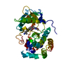
 Basic information
 Components
 Keywords
 Function and homology information
 Homo sapiens (human)
 X-RAY DIFFRACTION /
  Authors
 Citation
 Structure visualization
 Downloads & links
 Other downloads


 PDBj
PDBj


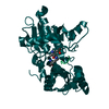
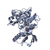
 Assembly
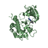

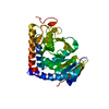
 Spodoptera frugiperda (fall armyworm)
 Mass: 326.312 Da / Num. of mol.: 2 / Source method: obtained synthetically / Formula: C18H10N6O
 Mass: 18.015 Da / Num. of mol.: 554 / Source method: isolated from a natural source / Formula: H2O
 Sample preparation
 / Beamline: X4C / Wavelength: 1 Å
 Processing