 Movie
Movie Controller


+ Open data

- Basic information
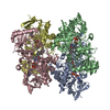
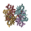
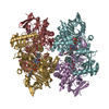
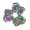
| Entry | Database: PDB / ID: 1ovl | ||||||
|---|---|---|---|---|---|---|---|
| Title | Crystal Structure of Nurr1 LBD | ||||||
 Components Components | (Orphan nuclear receptor NURR1 (MSE ...) x 2 | ||||||
 Keywords Keywords | TRANSCRIPTION / Nuur1 / LBD | ||||||
| Function / homology |  Function and homology information Function and homology informationgeneral adaptation syndrome / habenula development / cellular response to corticotropin-releasing hormone stimulus / central nervous system projection neuron axonogenesis / central nervous system neuron differentiation / nuclear glucocorticoid receptor binding / dopaminergic neuron differentiation / regulation of dopamine metabolic process / neuron maturation / midbrain dopaminergic neuron differentiation ...general adaptation syndrome / habenula development / cellular response to corticotropin-releasing hormone stimulus / central nervous system projection neuron axonogenesis / central nervous system neuron differentiation / nuclear glucocorticoid receptor binding / dopaminergic neuron differentiation / regulation of dopamine metabolic process / neuron maturation / midbrain dopaminergic neuron differentiation / regulation of respiratory gaseous exchange / dopamine biosynthetic process / canonical Wnt signaling pathway / negative regulation of apoptotic signaling pathway / fat cell differentiation / nuclear retinoid X receptor binding / adult locomotory behavior / post-embryonic development / response to amphetamine / SUMOylation of intracellular receptors / Nuclear Receptor transcription pathway / beta-catenin binding / neuron migration / nuclear receptor activity / sequence-specific double-stranded DNA binding / neuron apoptotic process / cellular response to oxidative stress / transcription by RNA polymerase II / DNA-binding transcription activator activity, RNA polymerase II-specific / transcription regulator complex / negative regulation of neuron apoptotic process / DNA-binding transcription factor activity, RNA polymerase II-specific / response to hypoxia / nuclear speck / RNA polymerase II cis-regulatory region sequence-specific DNA binding / protein heterodimerization activity / regulation of transcription by RNA polymerase II / chromatin / negative regulation of transcription by RNA polymerase II / DNA-templated transcription / positive regulation of transcription by RNA polymerase II / protein-containing complex / DNA binding / zinc ion binding / nucleoplasm / nucleus / cytoplasm Similarity search - Function | ||||||
| Biological species |  Homo sapiens (human) Homo sapiens (human) | ||||||
| Method |  X-RAY DIFFRACTION / SYNCHROTRON / MAD / Resolution: 2.2 Å X-RAY DIFFRACTION / SYNCHROTRON / MAD / Resolution: 2.2 Å | ||||||
 Authors Authors | Wang, Z. / Liu, J. / Walker, N. | ||||||
 Citation Citation | Journal: Nature / Year: 2003 Title: Structure and Function of Nurr1 identifies a Class of Ligand-Independent Nuclear Receptors Authors: Wang, Z. / Benoit, G. / Liu, J. / Prasad, S. / Aarnisalo, P. / Liu, X. / Xu, H. / Walker, N. / Perlmann, T. | ||||||
| History |
|
- Structure visualization
| Structure viewer | Molecule: MolmilJmol/JSmol |
|---|
- Downloads & links
-Download
| PDBx/mmCIF format | 1ovl.cif.gz | 295.2 KB | Display | PDBx/mmCIF format |
|---|---|---|---|---|
| PDB format | pdb1ovl.ent.gz | 237.7 KB | Display | PDB format |
| PDBx/mmJSON format | 1ovl.json.gz | Tree view | PDBx/mmJSON format | |
| Others |  Other downloads Other downloads |
-Validation report
| Arichive directory | https://data.pdbj.org/pub/pdb/validation_reports/ov/1ovlftp://data.pdbj.org/pub/pdb/validation_reports/ov/1ovl | HTTPS FTP |
|---|
-Related structure data
| Similar structure data |
|---|

-Links
 PDBj
PDBj

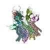
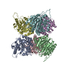
- Assembly
| Deposited unit | 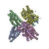
| ||||||||
|---|---|---|---|---|---|---|---|---|---|
| 1 |
| ||||||||
| Unit cell |
|
-Components
-Orphan nuclear receptor NURR1 (MSE ... , 2 types, 6 molecules ADBCEF
| #1: Protein | Mass: 30670.041 Da / Num. of mol.: 2 Source method: isolated from a genetically manipulated source Source: (gene. exp.) Homo sapiens (human) / Gene: NR4A2 OR NURR1 OR TINUR OR NOT / Plasmid: pET-15b / Production host:  #2: Protein | Mass: 30716.936 Da / Num. of mol.: 4 Source method: isolated from a genetically manipulated source Source: (gene. exp.) Homo sapiens (human) / Gene: NR4A2 OR NURR1 OR TINUR OR NOT / Plasmid: pET-15b / Production host: |
|---|
-Non-polymers , 4 types, 590 molecules 


| #3: Chemical | ChemComp-K /  Mass: 39.098 Da / Num. of mol.: 8 / Source method: obtained synthetically / Formula: K Mass: 39.098 Da / Num. of mol.: 8 / Source method: obtained synthetically / Formula: K#4: Chemical | ChemComp-BR /  Mass: 79.904 Da / Num. of mol.: 19 / Source method: obtained synthetically / Formula: Br Mass: 79.904 Da / Num. of mol.: 19 / Source method: obtained synthetically / Formula: Br#5: Chemical | ChemComp-IOD / |  Mass: 126.904 Da / Num. of mol.: 1 / Source method: obtained synthetically / Formula: I Mass: 126.904 Da / Num. of mol.: 1 / Source method: obtained synthetically / Formula: I#6: Water | ChemComp-HOH / | Mass: 18.015 Da / Num. of mol.: 562 / Source method: isolated from a natural source / Formula: H2O |
|---|
-Details
| Has protein modification | Y |
|---|
-Experimental details
-Experiment
| Experiment | Method: X-RAY DIFFRACTION / Number of used crystals: 1 |
|---|
- Sample preparation
| Crystal | Density Matthews: 2.3 Å3/Da / Density % sol: 46.54 % | |||||||||||||||||||||||||||||||||||||||||||||||||||||||||||||||
|---|---|---|---|---|---|---|---|---|---|---|---|---|---|---|---|---|---|---|---|---|---|---|---|---|---|---|---|---|---|---|---|---|---|---|---|---|---|---|---|---|---|---|---|---|---|---|---|---|---|---|---|---|---|---|---|---|---|---|---|---|---|---|---|---|
| Crystal grow | Temperature: 298 K / Method: vapor diffusion / pH: 6.5 Details: PEG3350,KBr, Hepes, pH 6.5, VAPOR DIFFUSION, temperature 298K | |||||||||||||||||||||||||||||||||||||||||||||||||||||||||||||||
| Crystal grow | *PLUS Temperature: 20 ℃ / pH: 7.9 / Method: vapor diffusion, hanging drop | |||||||||||||||||||||||||||||||||||||||||||||||||||||||||||||||
| Components of the solutions | *PLUS
|
-Data collection
| Diffraction | Mean temperature: 90 K |
|---|---|
| Diffraction source | Source: SYNCHROTRON / Site: ALS  / Beamline: 5.0.2 / Wavelength: 1.1 Å / Beamline: 5.0.2 / Wavelength: 1.1 Å |
| Detector | Type: ADSC QUANTUM 4 / Detector: CCD / Date: Nov 29, 2001 |
| Radiation | Monochromator: Double cyrstal Si(111) / Protocol: SINGLE WAVELENGTH / Monochromatic (M) / Laue (L): M / Scattering type: x-ray |
| Radiation wavelength | Wavelength: 1.1 Å / Relative weight: 1 |
| Reflection | Resolution: 2.2→34.7 Å / Num. all: 287222 / Num. obs: 82388 / % possible obs: 98.7 % / Observed criterion σ(F): 1 / Observed criterion σ(I): 1 / Rsym value: 0.072 / Net I/σ(I): 6.2 |
| Reflection shell | Resolution: 2.2→2.32 Å / Rmerge(I) obs: 0.269 / Mean I/σ(I) obs: 2.4 / % possible all: 91.7 |
| Reflection | *PLUS Num. measured all: 287222 / Rmerge(I) obs: 0.072 |
- Processing
| Software |
| |||||||||||||||||||||||||
|---|---|---|---|---|---|---|---|---|---|---|---|---|---|---|---|---|---|---|---|---|---|---|---|---|---|---|
| Refinement | Method to determine structure: MAD / Resolution: 2.2→500 Å / σ(F): 0 / Stereochemistry target values: Engh & Huber
| |||||||||||||||||||||||||
| Refinement step | Cycle: LAST / Resolution: 2.2→500 Å
| |||||||||||||||||||||||||
| Refine LS restraints |
| |||||||||||||||||||||||||
| Refinement | *PLUS % reflection Rfree: 5 % | |||||||||||||||||||||||||
| Solvent computation | *PLUS | |||||||||||||||||||||||||
| Displacement parameters | *PLUS |
