 Movie
Movie Controller


+ Open data

- Basic information
| Entry | Database: PDB / ID: 1cev | ||||||
|---|---|---|---|---|---|---|---|
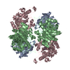
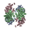
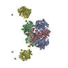
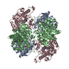
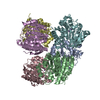
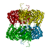
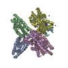
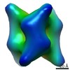
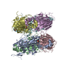
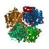
| Title | ARGINASE FROM BACILLUS CALDOVELOX, NATIVE STRUCTURE AT PH 5.6 | ||||||
 Components Components | PROTEIN (ARGINASE) | ||||||
 Keywords Keywords | HYDROLASE / ENZYME / ARGININE HYDROLYSIS / NITROGEN METABOLISM / MANGANESE METALLOENZYME | ||||||
| Function / homology |  Function and homology information Function and homology informationarginine metabolic process / arginase / arginase activity / urea cycle / manganese ion binding / cytoplasm Similarity search - Function | ||||||
| Biological species |  Bacillus caldovelox (bacteria) Bacillus caldovelox (bacteria) | ||||||
| Method |  X-RAY DIFFRACTION / MOLECULAR REPLACEMENT / Resolution: 2.4 Å X-RAY DIFFRACTION / MOLECULAR REPLACEMENT / Resolution: 2.4 Å | ||||||
 Authors Authors | Bewley, M.C. / Jeffrey, P.D. / Patchett, M.L. / Kanyo, Z.F. / Baker, E.N. | ||||||
 Citation Citation | Journal: Structure Fold.Des. / Year: 1999 Title: Crystal structures of Bacillus caldovelox arginase in complex with substrate and inhibitors reveal new insights into activation, inhibition and catalysis in the arginase superfamily. Authors: Bewley, M.C. / Jeffrey, P.D. / Patchett, M.L. / Kanyo, Z.F. / Baker, E.N. | ||||||
| History |
|
- Structure visualization
| Structure viewer | Molecule: MolmilJmol/JSmol |
|---|
- Downloads & links
-Download
| PDBx/mmCIF format | 1cev.cif.gz | 334.4 KB | Display | PDBx/mmCIF format |
|---|---|---|---|---|
| PDB format | pdb1cev.ent.gz | 276.6 KB | Display | PDB format |
| PDBx/mmJSON format | 1cev.json.gz | Tree view | PDBx/mmJSON format | |
| Others |  Other downloads Other downloads |
-Validation report
| Arichive directory | https://data.pdbj.org/pub/pdb/validation_reports/ce/1cevftp://data.pdbj.org/pub/pdb/validation_reports/ce/1cev | HTTPS FTP |
|---|
-Related structure data


-Links
 PDBj
PDBj- Assembly
| Deposited unit | 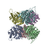
| ||||||||
|---|---|---|---|---|---|---|---|---|---|
| 1 |
| ||||||||
| 2 | 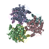
| ||||||||
| Unit cell |
|
-Components
| #1: Protein | Mass: 32476.248 Da / Num. of mol.: 6 Source method: isolated from a genetically manipulated source Source: (gene. exp.) Bacillus caldovelox (bacteria) / Strain: BACILLUS SPECIES DSM 411 / Description: BACILLUS SPECIES DSM 411 / Cell line (production host): BL21 / Production host: #2: Chemical | ChemComp-MN /   Mass: 54.938 Da / Num. of mol.: 12 / Source method: obtained synthetically / Formula: Mn Mass: 54.938 Da / Num. of mol.: 12 / Source method: obtained synthetically / Formula: Mn |
|---|
-Experimental details
-Experiment
| Experiment | Method: X-RAY DIFFRACTION / Number of used crystals: 1 |
|---|
- Sample preparation
| Crystal | Density Matthews: 2.46 Å3/Da / Density % sol: 50 % | |||||||||||||||||||||||||
|---|---|---|---|---|---|---|---|---|---|---|---|---|---|---|---|---|---|---|---|---|---|---|---|---|---|---|
| Crystal grow | Method: vapor diffusion, hanging drop / pH: 5.6 Details: 3-6% N-BUTANOL,IN 0.1 M SODIUM CITRATE BUFFER, PH 5.6 PROTEIN SOLUTION 27 MG/ML PROTEIN, 10 MM MOPS, PH 7.5, VAPOR DIFFUSION, HANGING DROP | |||||||||||||||||||||||||
| Crystal grow | *PLUS | |||||||||||||||||||||||||
| Components of the solutions | *PLUS
|
-Data collection
| Diffraction | Mean temperature: 113 K |
|---|---|
| Diffraction source | Source: ROTATING ANODE / Type: RIGAKU RU200 / Wavelength: 1.5418 |
| Detector | Type: RIGAKU / Detector: IMAGE PLATE / Date: Nov 1, 1996 |
| Radiation | Monochromator: GRAPHITE / Protocol: SINGLE WAVELENGTH / Monochromatic (M) / Laue (L): M / Scattering type: x-ray |
| Radiation wavelength | Wavelength: 1.5418 Å / Relative weight: 1 |
| Reflection | Resolution: 2.4→40 Å / Num. obs: 68531 / % possible obs: 90.9 % / Redundancy: 4.2 % / Rmerge(I) obs: 0.071 / Net I/σ(I): 15.8 |
| Reflection shell | Resolution: 2.4→2.6 Å / Rmerge(I) obs: 0.344 / Mean I/σ(I) obs: 2.8 / % possible all: 67.1 |
| Reflection | *PLUS Num. measured all: 286814 |
| Reflection shell | *PLUS % possible obs: 67.1 % |
- Processing
| Software |
| ||||||||||||||||||||||||||||||||||||||||||||||||||||||||||||
|---|---|---|---|---|---|---|---|---|---|---|---|---|---|---|---|---|---|---|---|---|---|---|---|---|---|---|---|---|---|---|---|---|---|---|---|---|---|---|---|---|---|---|---|---|---|---|---|---|---|---|---|---|---|---|---|---|---|---|---|---|---|
| Refinement | Method to determine structure: MOLECULAR REPLACEMENT Starting model: LOW RESOLUTION STRUCTURE OF A DIFFERENT CRYSTAL FORM Resolution: 2.4→6 Å / Isotropic thermal model: INDIVIDUAL ISOTROPIC / Cross valid method: THROUGHOUT / σ(F): 0
| ||||||||||||||||||||||||||||||||||||||||||||||||||||||||||||
| Refine analyze | Luzzati d res low obs: 6 Å | ||||||||||||||||||||||||||||||||||||||||||||||||||||||||||||
| Refinement step | Cycle: LAST / Resolution: 2.4→6 Å
| ||||||||||||||||||||||||||||||||||||||||||||||||||||||||||||
| Refine LS restraints |
| ||||||||||||||||||||||||||||||||||||||||||||||||||||||||||||
| Refine LS restraints NCS | NCS model details: RESTRAINTS | ||||||||||||||||||||||||||||||||||||||||||||||||||||||||||||
| Xplor file | Serial no: 1 / Param file: PARAM19X.PRO / Topol file: TOPH19X.PRO | ||||||||||||||||||||||||||||||||||||||||||||||||||||||||||||
| Software | *PLUS Name: X-PLOR / Version: 3.1 / Classification: refinement | ||||||||||||||||||||||||||||||||||||||||||||||||||||||||||||
| Refinement | *PLUS Rfactor obs: 0.205 | ||||||||||||||||||||||||||||||||||||||||||||||||||||||||||||
| Solvent computation | *PLUS | ||||||||||||||||||||||||||||||||||||||||||||||||||||||||||||
| Displacement parameters | *PLUS |
