 Movie
Movie Controller


+ Open data

- Basic information
| Entry | Database: PDB / ID: 1onh | ||||||
|---|---|---|---|---|---|---|---|
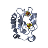
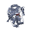
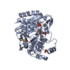
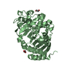
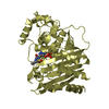
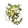
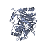
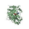
| Title | GC1 beta-lactamase with a penem inhibitor | ||||||
 Components Components | class C beta-lactamase | ||||||
 Keywords Keywords | HYDROLASE / MIXED ALPHA/BETA / CEPHALOSPORINASE / INHIBITION / CLASS C BETA-LACTAMASE | ||||||
| Function / homology |  Function and homology information Function and homology informationantibiotic catabolic process / beta-lactamase activity / beta-lactamase / outer membrane-bounded periplasmic space / response to antibiotic Similarity search - Function | ||||||
| Biological species |  Enterobacter cloacae (bacteria) Enterobacter cloacae (bacteria) | ||||||
| Method |  X-RAY DIFFRACTION / SYNCHROTRON / MOLECULAR REPLACEMENT / Resolution: 1.38 Å X-RAY DIFFRACTION / SYNCHROTRON / MOLECULAR REPLACEMENT / Resolution: 1.38 Å | ||||||
 Authors Authors | Nukaga, M. / Nukaga, K. / Knox, J.R. | ||||||
 Citation Citation | Journal: Biochemistry / Year: 2003 Title: Inhibition of Class A and Class C Beta-Lactamases by Penems: Crystallographic Structures of a Novel 1,4-Thiazepine Intermediate Authors: Nukaga, M. / Abe, T. / Venkatesan, A.M. / Mansour, T.S. / Bonomo, R.A. / Knox, J.R. | ||||||
| History |
|
- Structure visualization
| Structure viewer | Molecule: MolmilJmol/JSmol |
|---|
- Downloads & links
-Download
| PDBx/mmCIF format | 1onh.cif.gz | 175.5 KB | Display | PDBx/mmCIF format |
|---|---|---|---|---|
| PDB format | pdb1onh.ent.gz | 139.1 KB | Display | PDB format |
| PDBx/mmJSON format | 1onh.json.gz | Tree view | PDBx/mmJSON format | |
| Others |  Other downloads Other downloads |
-Validation report
| Arichive directory | https://data.pdbj.org/pub/pdb/validation_reports/on/1onhftp://data.pdbj.org/pub/pdb/validation_reports/on/1onh | HTTPS FTP |
|---|
-Related structure data
| Related structure data |  1ongC 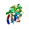 1gceS S: Starting model for refinement C: citing same article ( |
|---|---|
| Similar structure data |


-Links
 PDBj
PDBj


- Assembly
| Deposited unit | 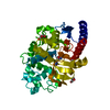
| |||||||||
|---|---|---|---|---|---|---|---|---|---|---|
| 1 |
| |||||||||
| Unit cell |
| |||||||||
| Components on special symmetry positions |
|
-Components
| #1: Protein | Mass: 39609.172 Da / Num. of mol.: 1 Source method: isolated from a genetically manipulated source Source: (gene. exp.) Enterobacter cloacae (bacteria) / Gene: BLAC / Plasmid: PCS101 / Production host: | ||||
|---|---|---|---|---|---|
| #2: Chemical | ChemComp-WY4 /   Mass: 307.325 Da / Num. of mol.: 1 / Source method: obtained synthetically / Formula: C13H13N3O4S Mass: 307.325 Da / Num. of mol.: 1 / Source method: obtained synthetically / Formula: C13H13N3O4S | ||||
| #3: Chemical | ChemComp-GOL /   Mass: 92.094 Da / Num. of mol.: 4 / Source method: obtained synthetically / Formula: C3H8O3 Mass: 92.094 Da / Num. of mol.: 4 / Source method: obtained synthetically / Formula: C3H8O3#4: Water | ChemComp-HOH / |  Mass: 18.015 Da / Num. of mol.: 427 / Source method: isolated from a natural source / Formula: H2O Mass: 18.015 Da / Num. of mol.: 427 / Source method: isolated from a natural source / Formula: H2OHas protein modification | Y | |
-Experimental details
-Experiment
| Experiment | Method: X-RAY DIFFRACTION / Number of used crystals: 1 |
|---|
- Sample preparation
| Crystal | Density Matthews: 2.17 Å3/Da / Density % sol: 43 % | ||||||||||||||||||||||||||||||||||||
|---|---|---|---|---|---|---|---|---|---|---|---|---|---|---|---|---|---|---|---|---|---|---|---|---|---|---|---|---|---|---|---|---|---|---|---|---|---|
| Crystal grow | Temperature: 293 K / Method: vapor diffusion, sitting drop / pH: 7 Details: 10% PEG 8000 in 50 mM HEPES over 20 % PEG, pH 7.0, VAPOR DIFFUSION, SITTING DROP, temperature 293K | ||||||||||||||||||||||||||||||||||||
| Crystal grow | *PLUS Method: vapor diffusion, sitting drop | ||||||||||||||||||||||||||||||||||||
| Components of the solutions | *PLUS
|
-Data collection
| Diffraction | Mean temperature: 100 K |
|---|---|
| Diffraction source | Source: SYNCHROTRON / Site: CHESS  / Beamline: A1 / Wavelength: 0.9474 / Wavelength: 0.9474 Å / Beamline: A1 / Wavelength: 0.9474 / Wavelength: 0.9474 Å |
| Detector | Type: ADSC QUANTUM 210 / Detector: CCD / Date: Jun 24, 2002 |
| Radiation | Protocol: SINGLE WAVELENGTH / Monochromatic (M) / Laue (L): M / Scattering type: x-ray |
| Radiation wavelength | Wavelength: 0.9474 Å / Relative weight: 1 |
| Reflection | Resolution: 1.38→50 Å / Num. obs: 65047 / % possible obs: 97.2 % / Observed criterion σ(F): 0 / Observed criterion σ(I): 0 / Redundancy: 5.8 % / Rmerge(I) obs: 0.103 / Net I/σ(I): 10.5 |
| Reflection shell | Resolution: 1.38→1.43 Å / Redundancy: 2.52 % / Rmerge(I) obs: 0.514 / Mean I/σ(I) obs: 2 / Num. unique all: 5075 / % possible all: 77 |
| Reflection | *PLUS Highest resolution: 1.38 Å / Num. measured all: 377696 |
| Reflection shell | *PLUS % possible obs: 77 % / Num. unique obs: 5075 / Num. measured obs: 12765 |
- Processing
| Software |
| |||||||||||||||||||||||||||||||||
|---|---|---|---|---|---|---|---|---|---|---|---|---|---|---|---|---|---|---|---|---|---|---|---|---|---|---|---|---|---|---|---|---|---|---|
| Refinement | Method to determine structure: MOLECULAR REPLACEMENT Starting model: PDB ENTRY 1GCE Resolution: 1.38→20 Å / Num. parameters: 30398 / Num. restraintsaints: 38104 / Isotropic thermal model: anisotropic / Cross valid method: THROUGHOUT / σ(F): 0 / Stereochemistry target values: Engh & Huber
| |||||||||||||||||||||||||||||||||
| Refine analyze | Num. disordered residues: 36 / Occupancy sum hydrogen: 2680.96 / Occupancy sum non hydrogen: 3219.6 | |||||||||||||||||||||||||||||||||
| Refinement step | Cycle: LAST / Resolution: 1.38→20 Å
| |||||||||||||||||||||||||||||||||
| Refine LS restraints |
| |||||||||||||||||||||||||||||||||
| Software | *PLUS Name: SHELXL / Version: 97 / Classification: refinement | |||||||||||||||||||||||||||||||||
| Refinement | *PLUS Lowest resolution: 20 Å / Rfactor Rfree: 0.202 / Rfactor Rwork: 0.138 | |||||||||||||||||||||||||||||||||
| Solvent computation | *PLUS | |||||||||||||||||||||||||||||||||
| Displacement parameters | *PLUS | |||||||||||||||||||||||||||||||||
| Refine LS restraints | *PLUS
|
