 Movie
Movie Controller

[English] 日本語
 Yorodumi
- PDB-1ofh: Asymmetric complex between HslV and I-domain deleted HslU (H. inf... -
+ Open data

- Basic information
| Entry | Database: PDB / ID: 1ofh | ||||||
|---|---|---|---|---|---|---|---|
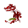
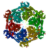
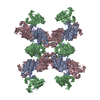
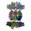
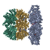
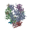
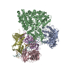
| Title | Asymmetric complex between HslV and I-domain deleted HslU (H. influenzae) | ||||||
 Components Components | (ATP-DEPENDENT ...) x 2 | ||||||
 Keywords Keywords | HYDROLASE / CHAPERONE / ATP-BINDING | ||||||
| Function / homology |  Function and homology information Function and homology informationHslU-HslV peptidase / HslUV protease complex / proteasome-activating activity / proteasome core complex / protein unfolding / threonine-type endopeptidase activity / : / peptidase activity / ATP hydrolysis activity / ATP binding ...HslU-HslV peptidase / HslUV protease complex / proteasome-activating activity / proteasome core complex / protein unfolding / threonine-type endopeptidase activity / : / peptidase activity / ATP hydrolysis activity / ATP binding / metal ion binding / cytoplasm Similarity search - Function | ||||||
| Biological species |  HAEMOPHILUS INFLUENZAE (bacteria) HAEMOPHILUS INFLUENZAE (bacteria) | ||||||
| Method |  X-RAY DIFFRACTION / SYNCHROTRON / MOLECULAR REPLACEMENT / Resolution: 2.5 Å X-RAY DIFFRACTION / SYNCHROTRON / MOLECULAR REPLACEMENT / Resolution: 2.5 Å | ||||||
 Authors Authors | Kwon, A.R. / Kessler, B.M. / Overkleeft, H.S. / McKay, D.B. | ||||||
 Citation Citation | Journal: J.Mol.Biol. / Year: 2003 Title: Structure and Reactivity of an Asymmetric Complex between Hslv and I-Domain Deleted Hslu, a Prokaryotic Homolog of the Eukaryotic Proteasome Authors: Kwon, A.R. / Kessler, B.M. / Overkleeft, H.S. / Mckay, D.B. #1: Journal: J.Mol.Biol. / Year: 2002Title: Structure of Hsluv Complexed with a Vinyl Sulfone Inhibitor: Corroboration of a Proposed Mechanism of Allosteric Activation of Hslv by Hslu Authors: Sousa, M.C. / Kessler, B.M. / Overkleeft, H.S. / Mckay, D.B. #2: Journal: Cell(Cambridge,Mass.) / Year: 2000Title: Crystal and Solution Structures of an Hsluv Protease-Chaperone Complex Authors: Sousa, M.C. / Trame, C.B. / Tsuruta, H. / Wilbanks, S.M. / Reddy, V.S. / Mckay, D.B. | ||||||
| History |
|
- Structure visualization
| Structure viewer | Molecule: MolmilJmol/JSmol |
|---|
- Downloads & links
-Download
| PDBx/mmCIF format | 1ofh.cif.gz | 382.9 KB | Display | PDBx/mmCIF format |
|---|---|---|---|---|
| PDB format | pdb1ofh.ent.gz | 311.5 KB | Display | PDB format |
| PDBx/mmJSON format | 1ofh.json.gz | Tree view | PDBx/mmJSON format | |
| Others |  Other downloads Other downloads |
-Validation report
| Arichive directory | https://data.pdbj.org/pub/pdb/validation_reports/of/1ofhftp://data.pdbj.org/pub/pdb/validation_reports/of/1ofh | HTTPS FTP |
|---|
-Related structure data
| Related structure data |  1ofiC 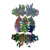 1g3iS S: Starting model for refinement C: citing same article ( |
|---|---|
| Similar structure data |


-Links
 PDBj
PDBj


- Assembly
| Deposited unit | 
| ||||||||
|---|---|---|---|---|---|---|---|---|---|
| 1 | x 6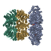
| ||||||||
| 2 | 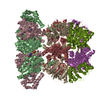
| ||||||||
| Unit cell |
|
-Components
-ATP-DEPENDENT ... , 2 types, 9 molecules ABCGHILMN
| #1: Protein | Mass: 34126.020 Da / Num. of mol.: 3 / Fragment: RESIDUES 1-107,244-444 Source method: isolated from a genetically manipulated source Source: (gene. exp.) HAEMOPHILUS INFLUENZAE (bacteria) / Strain: RD / Plasmid: PET / Production host: #2: Protein | Mass: 18903.549 Da / Num. of mol.: 6 Source method: isolated from a genetically manipulated source Source: (gene. exp.) HAEMOPHILUS INFLUENZAE (bacteria) / Strain: RD / Plasmid: PET / Production host: References: UniProt: P43772, Hydrolases; Acting on peptide bonds (peptidases); Threonine endopeptidases |
|---|
-Non-polymers , 4 types, 98 molecules 


| #3: Chemical |  Mass: 427.201 Da / Num. of mol.: 3 / Source method: obtained synthetically / Formula: C10H15N5O10P2 / Comment: ADP, energy-carrying molecule*YM Mass: 427.201 Da / Num. of mol.: 3 / Source method: obtained synthetically / Formula: C10H15N5O10P2 / Comment: ADP, energy-carrying molecule*YM#4: Chemical | ChemComp-MG /  Mass: 24.305 Da / Num. of mol.: 12 / Source method: obtained synthetically / Formula: Mg Mass: 24.305 Da / Num. of mol.: 12 / Source method: obtained synthetically / Formula: Mg#5: Chemical |  Mass: 94.971 Da / Num. of mol.: 3 / Source method: obtained synthetically / Formula: PO4 Mass: 94.971 Da / Num. of mol.: 3 / Source method: obtained synthetically / Formula: PO4#6: Water | ChemComp-HOH / | Mass: 18.015 Da / Num. of mol.: 80 / Source method: isolated from a natural source / Formula: H2O |
|---|
-Details
| Compound details | FUNCTION: CHAPERONE SUBUNIT OF A PROTEASOME |
|---|
-Experimental details
-Experiment
| Experiment | Method: X-RAY DIFFRACTION / Number of used crystals: 1 |
|---|
- Sample preparation
| Crystal | Density Matthews: 2.86 Å3/Da / Density % sol: 57 % | |||||||||||||||||||||||||||||||||||||||||||||||||||||||||||||||
|---|---|---|---|---|---|---|---|---|---|---|---|---|---|---|---|---|---|---|---|---|---|---|---|---|---|---|---|---|---|---|---|---|---|---|---|---|---|---|---|---|---|---|---|---|---|---|---|---|---|---|---|---|---|---|---|---|---|---|---|---|---|---|---|---|
| Crystal grow | pH: 5.5 / Details: PEG-MME 2000, KCL,MG(OAC)2,CITRATE, PH 5.5 | |||||||||||||||||||||||||||||||||||||||||||||||||||||||||||||||
| Crystal grow | *PLUS pH: 7 / Method: unknown | |||||||||||||||||||||||||||||||||||||||||||||||||||||||||||||||
| Components of the solutions | *PLUS
|
-Data collection
| Diffraction | Mean temperature: 100 K |
|---|---|
| Diffraction source | Source: SYNCHROTRON / Site: SSRL  / Beamline: BL11-1 / Wavelength: 0.98 / Beamline: BL11-1 / Wavelength: 0.98 |
| Detector | Type: ADSC CCD / Detector: CCD / Date: Mar 15, 2002 / Details: MIRRORS |
| Radiation | Monochromator: SI (111) / Protocol: SINGLE WAVELENGTH / Monochromatic (M) / Laue (L): M / Scattering type: x-ray |
| Radiation wavelength | Wavelength: 0.98 Å / Relative weight: 1 |
| Reflection | Resolution: 2.5→40 Å / Num. obs: 81671 / % possible obs: 97 % / Observed criterion σ(I): 0 / Redundancy: 4.2 % / Rmerge(I) obs: 0.073 / Net I/σ(I): 39.8 |
| Reflection shell | Resolution: 2.5→2.54 Å / Mean I/σ(I) obs: 2.3 / % possible all: 74.9 |
| Reflection | *PLUS Highest resolution: 2.5 Å / Lowest resolution: 40 Å / Num. measured all: 344637 / Rmerge(I) obs: 0.073 |
| Reflection shell | *PLUS % possible obs: 74.9 % / Rmerge(I) obs: 0.319 / Mean I/σ(I) obs: 2.3 |
- Processing
| Software |
| ||||||||||||||||||||||||||||||||||||||||||||||||||||||||||||
|---|---|---|---|---|---|---|---|---|---|---|---|---|---|---|---|---|---|---|---|---|---|---|---|---|---|---|---|---|---|---|---|---|---|---|---|---|---|---|---|---|---|---|---|---|---|---|---|---|---|---|---|---|---|---|---|---|---|---|---|---|---|
| Refinement | Method to determine structure: MOLECULAR REPLACEMENT Starting model: PDB ENTRY 1G3I Resolution: 2.5→40 Å / Cross valid method: THROUGHOUT / σ(F): 0
| ||||||||||||||||||||||||||||||||||||||||||||||||||||||||||||
| Solvent computation | Solvent model: DENSITY MODIFICATION | ||||||||||||||||||||||||||||||||||||||||||||||||||||||||||||
| Refine analyze | Luzzati coordinate error obs: 0.37 Å | ||||||||||||||||||||||||||||||||||||||||||||||||||||||||||||
| Refinement step | Cycle: LAST / Resolution: 2.5→40 Å
| ||||||||||||||||||||||||||||||||||||||||||||||||||||||||||||
| Refine LS restraints |
| ||||||||||||||||||||||||||||||||||||||||||||||||||||||||||||
| LS refinement shell | Resolution: 2.5→2.52 Å / Total num. of bins used: 20
| ||||||||||||||||||||||||||||||||||||||||||||||||||||||||||||
| Refinement | *PLUS Num. reflection obs: 71454 | ||||||||||||||||||||||||||||||||||||||||||||||||||||||||||||
| Solvent computation | *PLUS | ||||||||||||||||||||||||||||||||||||||||||||||||||||||||||||
| Displacement parameters | *PLUS |
