 Movie
Movie Controller

[English] 日本語
 Yorodumi
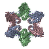
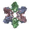
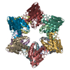
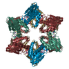
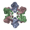
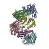
- PDB-1jjw: Structure of Haemophilus influenzae HslV Protein at 1.9 A Resolution -
+ Open data

- Basic information
| Entry | Database: PDB / ID: 1jjw | ||||||
|---|---|---|---|---|---|---|---|
| Title | Structure of Haemophilus influenzae HslV Protein at 1.9 A Resolution | ||||||
 Components Components | ATP-DEPENDENT PROTEASE HSLV | ||||||
 Keywords Keywords | HYDROLASE / K anomalous scattering / quasi-equivalent packing | ||||||
| Function / homology |  Function and homology information Function and homology informationHslU-HslV peptidase / HslUV protease complex / proteasome core complex / threonine-type endopeptidase activity / : / metal ion binding / cytoplasm Similarity search - Function | ||||||
| Biological species |  Haemophilus influenzae (bacteria) Haemophilus influenzae (bacteria) | ||||||
| Method |  X-RAY DIFFRACTION / SYNCHROTRON / MOLECULAR REPLACEMENT / Resolution: 1.9 Å X-RAY DIFFRACTION / SYNCHROTRON / MOLECULAR REPLACEMENT / Resolution: 1.9 Å | ||||||
 Authors Authors | Sousa, M.C. / McKay, D.B. | ||||||
 Citation Citation | Journal: Acta Crystallogr.,Sect.D / Year: 2001 Title: Structure of Haemophilus influenzae HslV protein at 1.9 A resolution, revealing a cation-binding site near the catalytic site. Authors: Sousa, M.C. / McKay, D.B. | ||||||
| History |
|
- Structure visualization
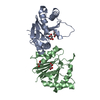
| Structure viewer | Molecule: MolmilJmol/JSmol |
|---|
- Downloads & links
-Download
| PDBx/mmCIF format | 1jjw.cif.gz | 110.8 KB | Display | PDBx/mmCIF format |
|---|---|---|---|---|
| PDB format | pdb1jjw.ent.gz | 85.4 KB | Display | PDB format |
| PDBx/mmJSON format | 1jjw.json.gz | Tree view | PDBx/mmJSON format | |
| Others |  Other downloads Other downloads |
-Validation report
| Arichive directory | https://data.pdbj.org/pub/pdb/validation_reports/jj/1jjwftp://data.pdbj.org/pub/pdb/validation_reports/jj/1jjw | HTTPS FTP |
|---|
-Related structure data
| Related structure data |  1g3kS S: Starting model for refinement |
|---|---|
| Similar structure data |

-Links
 PDBj
PDBj


- Assembly
| Deposited unit | 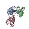
| ||||||||
|---|---|---|---|---|---|---|---|---|---|
| 1 | 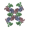
| ||||||||
| Unit cell |
|
-Components
| #1: Protein | Mass: 18903.549 Da / Num. of mol.: 3 Source method: isolated from a genetically manipulated source Source: (gene. exp.) Haemophilus influenzae (bacteria) / Gene: HslV / Plasmid: pRSET / Species (production host): Escherichia coli / Production host: #2: Chemical |   Mass: 39.098 Da / Num. of mol.: 3 / Source method: obtained synthetically / Formula: K Mass: 39.098 Da / Num. of mol.: 3 / Source method: obtained synthetically / Formula: K#3: Water | ChemComp-HOH / |  Mass: 18.015 Da / Num. of mol.: 191 / Source method: isolated from a natural source / Formula: H2O Mass: 18.015 Da / Num. of mol.: 191 / Source method: isolated from a natural source / Formula: H2O |
|---|
-Experimental details
-Experiment
| Experiment | Method: X-RAY DIFFRACTION / Number of used crystals: 1 |
|---|
- Sample preparation
| Crystal | Density Matthews: 2.69 Å3/Da / Density % sol: 54.21 % | ||||||||||||||||||||||||||||||||||||||||||||||||||||||||
|---|---|---|---|---|---|---|---|---|---|---|---|---|---|---|---|---|---|---|---|---|---|---|---|---|---|---|---|---|---|---|---|---|---|---|---|---|---|---|---|---|---|---|---|---|---|---|---|---|---|---|---|---|---|---|---|---|---|
| Crystal grow | Temperature: 277 K / Method: vapor diffusion, hanging drop / pH: 8.5 Details: sodium citrate, HCl Tris, PEG 400, pH 8.5, VAPOR DIFFUSION, HANGING DROP, temperature 277K | ||||||||||||||||||||||||||||||||||||||||||||||||||||||||
| Crystal grow | *PLUS pH: 7 | ||||||||||||||||||||||||||||||||||||||||||||||||||||||||
| Components of the solutions | *PLUS
|
-Data collection
| Diffraction | Mean temperature: 100 K |
|---|---|
| Diffraction source | Source: SYNCHROTRON / Site: SSRL  / Beamline: BL9-2 / Wavelength: 1.55 Å / Beamline: BL9-2 / Wavelength: 1.55 Å |
| Detector | Type: ADSC QUANTUM 4 / Detector: CCD / Date: Jan 6, 2000 |
| Radiation | Protocol: SINGLE WAVELENGTH / Monochromatic (M) / Laue (L): M / Scattering type: x-ray |
| Radiation wavelength | Wavelength: 1.55 Å / Relative weight: 1 |
| Reflection | Resolution: 1.9→30 Å / Num. all: 46472 / Num. obs: 46472 / % possible obs: 96.4 % / Observed criterion σ(F): 0 / Observed criterion σ(I): 0 / Biso Wilson estimate: 21.8 Å2 / Rmerge(I) obs: 0.049 / Rsym value: 0.049 / Net I/σ(I): 28.8 |
| Reflection shell | Resolution: 1.9→1.97 Å / Rmerge(I) obs: 0.195 / Mean I/σ(I) obs: 9.1 / Rsym value: 0.195 / % possible all: 98.3 |
- Processing
| Software |
| ||||||||||||||||||||
|---|---|---|---|---|---|---|---|---|---|---|---|---|---|---|---|---|---|---|---|---|---|
| Refinement | Method to determine structure: MOLECULAR REPLACEMENT Starting model: PDB ENTRY 1G3K Resolution: 1.9→19.9 Å / Rfactor Rfree error: 0.003 / Data cutoff high absF: 3273928.1 / Data cutoff low absF: 0 / Isotropic thermal model: RESTRAINED / Cross valid method: THROUGHOUT / σ(F): 0 / Stereochemistry target values: Engh & Huber
| ||||||||||||||||||||
| Solvent computation | Solvent model: FLAT MODEL / Bsol: 42.2708 Å2 / ksol: 0.36033 e/Å3 | ||||||||||||||||||||
| Displacement parameters | Biso mean: 33.7 Å2
| ||||||||||||||||||||
| Refine analyze |
| ||||||||||||||||||||
| Refinement step | Cycle: LAST / Resolution: 1.9→19.9 Å
| ||||||||||||||||||||
| Refine LS restraints |
| ||||||||||||||||||||
| LS refinement shell | Resolution: 1.9→2.02 Å / Rfactor Rfree error: 0.008 / Total num. of bins used: 6
| ||||||||||||||||||||
| Xplor file |
| ||||||||||||||||||||
| Software | *PLUS Name: CNS / Version: 1 / Classification: refinement | ||||||||||||||||||||
| Refinement | *PLUS Lowest resolution: 30 Å / σ(F): 0 / % reflection Rfree: 9.8 % / Rfactor obs: 0.217 / Rfactor Rfree: 0.251 | ||||||||||||||||||||
| Solvent computation | *PLUS | ||||||||||||||||||||
| Displacement parameters | *PLUS Biso mean: 33.7 Å2 | ||||||||||||||||||||
| Refine LS restraints | *PLUS
| ||||||||||||||||||||
| LS refinement shell | *PLUS Lowest resolution: 1.97 Å / Rfactor Rfree: 0.334 / % reflection Rfree: 9.8 % / Rfactor Rwork: 0.254 / Rfactor obs: 0.258 |
