Movie
Movie Controller


+ Open data

- Basic information
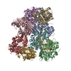
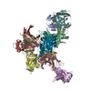
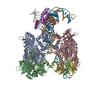
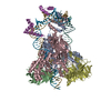
| Entry | Database: PDB / ID: 6knc | ||||||||||||||||||||||||
|---|---|---|---|---|---|---|---|---|---|---|---|---|---|---|---|---|---|---|---|---|---|---|---|---|---|
| Title | PolD-PCNA-DNA (form B) | ||||||||||||||||||||||||
 Components Components |
| ||||||||||||||||||||||||
 Keywords Keywords | REPLICATION/DNA / Family D Polymerase / PCNA / Switch Hook / REPLICATION / REPLICATION-DNA complex | ||||||||||||||||||||||||
| Function / homology |  Function and homology information Function and homology informationexodeoxyribonuclease I / DNA polymerase complex / intron homing / intein-mediated protein splicing / single-stranded DNA 3'-5' DNA exonuclease activity / DNA catabolic process / DNA strand elongation involved in DNA replication / leading strand elongation / DNA polymerase processivity factor activity / regulation of DNA replication ...exodeoxyribonuclease I / DNA polymerase complex / intron homing / intein-mediated protein splicing / single-stranded DNA 3'-5' DNA exonuclease activity / DNA catabolic process / DNA strand elongation involved in DNA replication / leading strand elongation / DNA polymerase processivity factor activity / regulation of DNA replication / DNA-templated DNA replication / endonuclease activity / DNA-directed DNA polymerase / DNA-directed DNA polymerase activity / DNA binding / metal ion binding / identical protein binding Similarity search - Function | ||||||||||||||||||||||||
| Biological species |   Thermococcus kodakarensis KOD1 (archaea)Thermococcus kodakarensis (archaea) Thermococcus kodakarensis KOD1 (archaea)Thermococcus kodakarensis (archaea)synthetic construct (others) | ||||||||||||||||||||||||
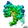
| Method | ELECTRON MICROSCOPY / single particle reconstruction / cryo EM / Resolution: 9.3 Å | ||||||||||||||||||||||||
 Authors Authors | Mayanagi, K. / Oki, K. / Miyazaki, N. / Ishino, S. / Yamagami, T. / Iwasaki, K. / Kohda, D. / Morikawa, K. / Shirai, T. / Ishino, Y. | ||||||||||||||||||||||||
| Funding support |  Japan, 7items Japan, 7items
| ||||||||||||||||||||||||
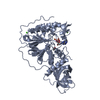
 Citation Citation | Journal: BMC Biol / Year: 2020 Title: Two conformations of DNA polymerase D-PCNA-DNA, an archaeal replisome complex, revealed by cryo-electron microscopy. Authors: Kouta Mayanagi / Keisuke Oki / Naoyuki Miyazaki / Sonoko Ishino / Takeshi Yamagami / Kosuke Morikawa / Kenji Iwasaki / Daisuke Kohda / Tsuyoshi Shirai / Yoshizumi Ishino / Abstract: BACKGROUND: DNA polymerase D (PolD) is the representative member of the D family of DNA polymerases. It is an archaea-specific DNA polymerase required for replication and unrelated to other known DNA ...BACKGROUND: DNA polymerase D (PolD) is the representative member of the D family of DNA polymerases. It is an archaea-specific DNA polymerase required for replication and unrelated to other known DNA polymerases. PolD consists of a heterodimer of two subunits, DP1 and DP2, which contain catalytic sites for 3'-5' editing exonuclease and DNA polymerase activities, respectively, with both proteins being mutually required for the full activities of each enzyme. However, the processivity of the replicase holoenzyme has additionally been shown to be enhanced by the clamp molecule proliferating cell nuclear antigen (PCNA), making it crucial to elucidate the interaction between PolD and PCNA on a structural level for a full understanding of its functional relevance. We present here the 3D structure of a PolD-PCNA-DNA complex from Thermococcus kodakarensis using single-particle cryo-electron microscopy (EM). RESULTS: Two distinct forms of the PolD-PCNA-DNA complex were identified by 3D classification analysis. Fitting the reported crystal structures of truncated forms of DP1 and DP2 from Pyrococcus ...RESULTS: Two distinct forms of the PolD-PCNA-DNA complex were identified by 3D classification analysis. Fitting the reported crystal structures of truncated forms of DP1 and DP2 from Pyrococcus abyssi onto our EM map showed the 3D atomic structural model of PolD-PCNA-DNA. In addition to the canonical interaction between PCNA and PolD via PIP (PCNA-interacting protein)-box motif, we found a new contact point consisting of a glutamate residue at position 171 in a β-hairpin of PCNA, which mediates interactions with DP1 and DP2. The DNA synthesis activity of a mutant PolD with disruption of the E171-mediated PCNA interaction was not stimulated by PCNA in vitro. CONCLUSIONS: Based on our analyses, we propose that glutamate residues at position 171 in each subunit of the PCNA homotrimer ring can function as hooks to lock PolD conformation on PCNA for ...CONCLUSIONS: Based on our analyses, we propose that glutamate residues at position 171 in each subunit of the PCNA homotrimer ring can function as hooks to lock PolD conformation on PCNA for conversion of its activity. This hook function of the clamp molecule may be conserved in the three domains of life. | ||||||||||||||||||||||||
| History |
|
- Structure visualization
| Movie |
Movie viewer |
|---|---|
| Structure viewer | Molecule: MolmilJmol/JSmol |
- Downloads & links
-Download
| PDBx/mmCIF format | 6knc.cif.gz | 460.8 KB | Display | PDBx/mmCIF format |
|---|---|---|---|---|
| PDB format | pdb6knc.ent.gz | 357.7 KB | Display | PDB format |
| PDBx/mmJSON format | 6knc.json.gz | Tree view | PDBx/mmJSON format | |
| Others |  Other downloads Other downloads |
-Validation report
| Arichive directory | https://data.pdbj.org/pub/pdb/validation_reports/kn/6kncftp://data.pdbj.org/pub/pdb/validation_reports/kn/6knc | HTTPS FTP |
|---|
-Related structure data
| Related structure data | 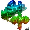 0725MC 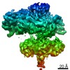 0723C 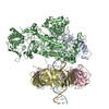 6knbC M: map data used to model this data C: citing same article ( |
|---|---|
| Similar structure data |


-Links
 PDBj
PDBj


- Assembly
| Deposited unit | 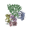
|
|---|---|
| 1 |
|
-Components
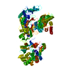
-DNA polymerase ... , 3 types, 5 molecules ABCDE
| #1: Protein | Mass: 60772.336 Da / Num. of mol.: 1 Source method: isolated from a genetically manipulated source Source: (gene. exp.) Thermococcus kodakarensis KOD1 (archaea)Strain: ATCC BAA-918 / JCM 12380 / KOD1 / Gene: polB, TK1902 / Production host:  References: UniProt: Q5JET1, DNA-directed DNA polymerase, exodeoxyribonuclease I |
|---|---|
| #2: Protein | Mass: 150418.047 Da / Num. of mol.: 1 Source method: isolated from a genetically manipulated source Source: (gene. exp.) Thermococcus kodakarensis (archaea) / Production host: |
| #3: Protein | Mass: 28270.441 Da / Num. of mol.: 3 Source method: isolated from a genetically manipulated source Source: (gene. exp.) Thermococcus kodakarensis KOD1 (archaea)Strain: ATCC BAA-918 / JCM 12380 / KOD1 / Gene: pcn1, TK0535 / Production host: |
-DNA chain , 2 types, 2 molecules FG
| #4: DNA chain | Mass: 9232.954 Da / Num. of mol.: 1 / Source method: obtained synthetically / Source: (synth.) synthetic construct (others) |
|---|---|
| #5: DNA chain | Mass: 9214.928 Da / Num. of mol.: 1 / Source method: obtained synthetically / Source: (synth.) synthetic construct (others) |
-Non-polymers , 2 types, 5 molecules 

| #6: Chemical | ChemComp-FE /  Mass: 55.845 Da / Num. of mol.: 1 / Source method: obtained synthetically / Formula: Fe Mass: 55.845 Da / Num. of mol.: 1 / Source method: obtained synthetically / Formula: Fe |
|---|---|
| #7: Chemical | ChemComp-ZN /  Mass: 65.409 Da / Num. of mol.: 4 / Source method: obtained synthetically / Formula: Zn Mass: 65.409 Da / Num. of mol.: 4 / Source method: obtained synthetically / Formula: Zn |
-Details
| Has ligand of interest | N |
|---|
-Experimental details
-Experiment
| Experiment | Method: ELECTRON MICROSCOPY |
|---|---|
| EM experiment | Aggregation state: PARTICLE / 3D reconstruction method: single particle reconstruction |
- Sample preparation
| Component | Name: Thermococcus kodakarensis PolD-PCNA-DNA / Type: COMPLEX / Entity ID: #1-#5 / Source: RECOMBINANT |
|---|---|
| Molecular weight | Value: 0.340 MDa / Experimental value: NO |
| Source (natural) | Organism: Thermococcus kodakarensis (archaea) |
| Source (recombinant) | Organism: |
| Buffer solution | pH: 8 |
| Specimen | Embedding applied: NO / Shadowing applied: NO / Staining applied: NO / Vitrification applied: YES |
| Specimen support | Grid material: GOLD / Grid type: Quantifoil R1.2/1.3 |
| Vitrification | Cryogen name: ETHANE |
- Electron microscopy imaging
| Experimental equipment |  Model: Titan Krios / Image courtesy: FEI Company |
|---|---|
| Microscopy | Model: FEI TITAN KRIOS |
| Electron gun | Electron source:  FIELD EMISSION GUN / Accelerating voltage: 300 kV / Illumination mode: FLOOD BEAM FIELD EMISSION GUN / Accelerating voltage: 300 kV / Illumination mode: FLOOD BEAM |
| Electron lens | Mode: BRIGHT FIELD |
| Image recording | Electron dose: 36 e/Å2 / Detector mode: INTEGRATING / Film or detector model: FEI FALCON II (4k x 4k) |
| EM imaging optics | Phase plate: VOLTA PHASE PLATE Spherical aberration corrector: Spherical aberration corrector |
- Processing
| Software | Name: PHENIX / Version: (1.11.1_2575: phenix.real_space_refine) / Classification: refinement | ||||||||||||||||||||||||
|---|---|---|---|---|---|---|---|---|---|---|---|---|---|---|---|---|---|---|---|---|---|---|---|---|---|
| EM software |
| ||||||||||||||||||||||||
| CTF correction | Type: PHASE FLIPPING AND AMPLITUDE CORRECTION | ||||||||||||||||||||||||
| Particle selection | Num. of particles selected: 240256 | ||||||||||||||||||||||||
| Symmetry | Point symmetry: C1 (asymmetric) | ||||||||||||||||||||||||
| 3D reconstruction | Resolution: 9.3 Å / Resolution method: FSC 0.143 CUT-OFF / Num. of particles: 19356 / Symmetry type: POINT | ||||||||||||||||||||||||
| Atomic model building | Protocol: FLEXIBLE FIT / Space: REAL / Target criteria: Correlation coefficient | ||||||||||||||||||||||||
| Atomic model building | 3D fitting-ID: 1 / Pdb chain-ID: A / Source name: PDB / Type: experimental model
| ||||||||||||||||||||||||
| Refinement | Stereochemistry target values: GeoStd + Monomer Library + CDL v1.2 | ||||||||||||||||||||||||
| Displacement parameters | Biso mean: 438.08 Å2 | ||||||||||||||||||||||||
| Refine LS restraints |
|