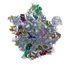
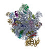
- EMDB-10074: SecA in complex with ribosome nascent chain -
+
Open data
ID or keywords:
Loading...
-
Basic information
Entry
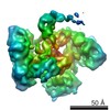
Database: EMDB / ID: EMD-10074
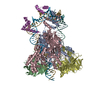
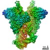
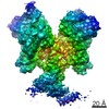
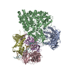
Title
SecA in complex with ribosome nascent chain
Map data
SecA bound to translating ribosome (with focused refined)
Sample
Complex: ribosome nascent chain in complex with SecA
Protein or peptide: SecA
Function / homology
Function and homology information
cell envelope Sec protein transport complex / protein-exporting ATPase activity / protein-secreting ATPase / protein transport by the Sec complex / intracellular protein transmembrane transport / protein import / protein targeting / ATP binding / metal ion binding / plasma membrane / cytosol Similarity search - Function
National Institutes of Health/National Center for Research Resources
United States
Citation
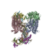
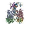
Journal: Nat Struct Mol Biol / Year: 2019 Title: The molecular mechanism of cotranslational membrane protein recognition and targeting by SecA. Authors: Shuai Wang / Ahmad Jomaa / Mateusz Jaskolowski / Chien-I Yang / Nenad Ban / Shu-Ou Shan / Abstract: Cotranslational protein targeting is a conserved process for membrane protein biogenesis. In Escherichia coli, the essential ATPase SecA was found to cotranslationally target a subset of nascent ...Cotranslational protein targeting is a conserved process for membrane protein biogenesis. In Escherichia coli, the essential ATPase SecA was found to cotranslationally target a subset of nascent membrane proteins to the SecYEG translocase at the plasma membrane. The molecular mechanism of this pathway remains unclear. Here we use biochemical and cryoelectron microscopy analyses to show that the amino-terminal amphipathic helix of SecA and the ribosomal protein uL23 form a composite binding site for the transmembrane domain (TMD) on the nascent protein. This binding mode further enables recognition of charged residues flanking the nascent TMD and thus explains the specificity of SecA recognition. Finally, we show that membrane-embedded SecYEG promotes handover of the translating ribosome from SecA to the translocase via a concerted mechanism. Our work provides a molecular description of the SecA-mediated cotranslational targeting pathway and demonstrates an unprecedented role of the ribosome in shielding nascent TMDs.
History
Deposition
Jun 17, 2019
-
Header (metadata) release
Oct 9, 2019
-
Map release
Oct 9, 2019
-
Update
Nov 27, 2019
-
Current status
Nov 27, 2019
Processing site: PDBe / Status: Released
-
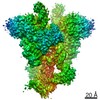
Structure visualization
Movie
Surface view with section colored by density value
In the structure databanks used in Yorodumi, some data are registered as the other names, "COVID-19 virus" and "2019-nCoV". Here are the details of the virus and the list of structure data.
Jan 31, 2019. EMDB accession codes are about to change! (news from PDBe EMDB page)
EMDB accession codes are about to change! (news from PDBe EMDB page)
The allocation of 4 digits for EMDB accession codes will soon come to an end. Whilst these codes will remain in use, new EMDB accession codes will include an additional digit and will expand incrementally as the available range of codes is exhausted. The current 4-digit format prefixed with “EMD-” (i.e. EMD-XXXX) will advance to a 5-digit format (i.e. EMD-XXXXX), and so on. It is currently estimated that the 4-digit codes will be depleted around Spring 2019, at which point the 5-digit format will come into force.
The EM Navigator/Yorodumi systems omit the EMD- prefix.
Related info.:Q: What is EMD? / ID/Accession-code notation in Yorodumi/EM Navigator
Yorodumi is a browser for structure data from EMDB, PDB, SASBDB, etc.
This page is also the successor to EM Navigator detail page, and also detail information page/front-end page for Omokage search.
The word "yorodu" (or yorozu) is an old Japanese word meaning "ten thousand". "mi" (miru) is to see.
Related info.:EMDB / PDB / SASBDB / Comparison of 3 databanks / Yorodumi Search / Aug 31, 2016. New EM Navigator & Yorodumi / Yorodumi Papers / Jmol/JSmol / Function and homology information / Changes in new EM Navigator and Yorodumi
 Movie
Movie Controller


 Open data
 Basic information
 Map data
 Sample
 Function and homology information
 Authors
 Switzerland,
  United States, 2 items
  Citation
 Structure visualization
 Downloads & links
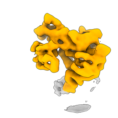 emd_10074.png
 http://ftp.pdbj.org/pub/emdb/structures/EMD-10074

 Z (Sec.)
 Y (Row.)
 X (Col.)


 Sample components
 Processing
 Electron microscopy
 FIELD EMISSION GUN