 Movie
Movie Controller


+ Open data

- Basic information
| Entry | Database: PDB / ID: 1g3i | ||||||
|---|---|---|---|---|---|---|---|
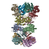
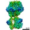
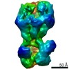
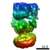
| Title | CRYSTAL STRUCTURE OF THE HSLUV PROTEASE-CHAPERONE COMPLEX | ||||||
 Components Components |
| ||||||
 Keywords Keywords | CHAPERONE/HYDROLASE / CHAPERONE-HYDROLASE complex | ||||||
| Function / homology |  Function and homology information Function and homology informationHslU-HslV peptidase / HslUV protease complex / proteasome-activating activity / proteasome core complex / protein unfolding / threonine-type endopeptidase activity / : / peptidase activity / ATP hydrolysis activity / ATP binding ...HslU-HslV peptidase / HslUV protease complex / proteasome-activating activity / proteasome core complex / protein unfolding / threonine-type endopeptidase activity / : / peptidase activity / ATP hydrolysis activity / ATP binding / metal ion binding / cytoplasm Similarity search - Function | ||||||
| Biological species |  Haemophilus influenzae (bacteria) Haemophilus influenzae (bacteria) | ||||||
| Method |  X-RAY DIFFRACTION / SYNCHROTRON / MOLECULAR REPLACEMENT / Resolution: 3.41 Å X-RAY DIFFRACTION / SYNCHROTRON / MOLECULAR REPLACEMENT / Resolution: 3.41 Å | ||||||
 Authors Authors | Sousa, M.C. / Trame, C.B. / Tsuruta, H. / Wilbanks, S.M. / Reddy, V.S. / McKay, D.B. | ||||||
 Citation Citation | Journal: Cell(Cambridge,Mass.) / Year: 2000 Title: Crystal and solution structures of an HslUV protease-chaperone complex. Authors: Sousa, M.C. / Trame, C.B. / Tsuruta, H. / Wilbanks, S.M. / Reddy, V.S. / McKay, D.B. | ||||||
| History |
|
- Structure visualization
| Structure viewer | Molecule: MolmilJmol/JSmol |
|---|
- Downloads & links
-Download
| PDBx/mmCIF format | 1g3i.cif.gz | 1.1 MB | Display | PDBx/mmCIF format |
|---|---|---|---|---|
| PDB format | pdb1g3i.ent.gz | 910.8 KB | Display | PDB format |
| PDBx/mmJSON format | 1g3i.json.gz | Tree view | PDBx/mmJSON format | |
| Others |  Other downloads Other downloads |
-Validation report
| Arichive directory | https://data.pdbj.org/pub/pdb/validation_reports/g3/1g3iftp://data.pdbj.org/pub/pdb/validation_reports/g3/1g3i | HTTPS FTP |
|---|
-Related structure data


-Links
 PDBj
PDBj


- Assembly
| Deposited unit | 
| ||||||||
|---|---|---|---|---|---|---|---|---|---|
| 1 |
| ||||||||
| Unit cell |
| ||||||||
| Details | The biological assembly is the complex HslU12-HslV12 seen in the asymmetric unit. |
-Components
| #1: Protein | Mass: 49441.504 Da / Num. of mol.: 12 Source method: isolated from a genetically manipulated source Source: (gene. exp.) Haemophilus influenzae (bacteria) / Species (production host): Escherichia coli / Production host: #2: Protein | Mass: 18903.549 Da / Num. of mol.: 12 Source method: isolated from a genetically manipulated source Source: (gene. exp.) Haemophilus influenzae (bacteria) / Species (production host): Escherichia coli / Production host: #3: Chemical | ChemComp-ATP /   Mass: 507.181 Da / Num. of mol.: 12 / Source method: obtained synthetically / Formula: C10H16N5O13P3 / Comment: ATP, energy-carrying molecule*YM Mass: 507.181 Da / Num. of mol.: 12 / Source method: obtained synthetically / Formula: C10H16N5O13P3 / Comment: ATP, energy-carrying molecule*YM |
|---|
-Experimental details
-Experiment
| Experiment | Method: X-RAY DIFFRACTION / Number of used crystals: 1 |
|---|
- Sample preparation
| Crystal | Density Matthews: 3.39 Å3/Da / Density % sol: 63.72 % | ||||||||||||||||||||||||||||||||||||||||||||||||||||||
|---|---|---|---|---|---|---|---|---|---|---|---|---|---|---|---|---|---|---|---|---|---|---|---|---|---|---|---|---|---|---|---|---|---|---|---|---|---|---|---|---|---|---|---|---|---|---|---|---|---|---|---|---|---|---|---|
| Crystal grow | Temperature: 298 K / Method: vapor diffusion, hanging drop / pH: 6 Details: PEG monomethyl ether 2000, potassium Chloride, magnesium acetate, citrate, pH 6.0, VAPOR DIFFUSION, HANGING DROP, temperature 298K | ||||||||||||||||||||||||||||||||||||||||||||||||||||||
| Crystal grow | *PLUS Temperature: 4 ℃ / pH: 7 | ||||||||||||||||||||||||||||||||||||||||||||||||||||||
| Components of the solutions | *PLUS
|
-Data collection
| Diffraction | Mean temperature: 100 K |
|---|---|
| Diffraction source | Source: SYNCHROTRON / Site: ALS  / Beamline: 5.0.2 / Wavelength: 1.1 Å / Beamline: 5.0.2 / Wavelength: 1.1 Å |
| Detector | Type: ADSC QUANTUM 4 / Detector: CCD / Date: Jun 23, 2000 |
| Radiation | Protocol: SINGLE WAVELENGTH / Monochromatic (M) / Laue (L): M / Scattering type: x-ray |
| Radiation wavelength | Wavelength: 1.1 Å / Relative weight: 1 |
| Reflection | Resolution: 3.4→30 Å / Num. all: 134912 / Num. obs: 134912 / % possible obs: 89.1 % / Observed criterion σ(F): 0 / Observed criterion σ(I): 0 / Redundancy: 2.2 % / Rmerge(I) obs: 0.064 / Net I/σ(I): 11.5 |
| Reflection shell | Resolution: 3.4→3.52 Å / Redundancy: 1.8 % / Rmerge(I) obs: 0.285 / Mean I/σ(I) obs: 3 / % possible all: 80.6 |
| Reflection | *PLUS Highest resolution: 3.4 Å / Lowest resolution: 30 Å / Num. measured all: 297864 |
| Reflection shell | *PLUS Highest resolution: 3.4 Å / % possible obs: 80.6 % / Mean I/σ(I) obs: 3 |
- Processing
| Software |
| |||||||||||||||||||||||||
|---|---|---|---|---|---|---|---|---|---|---|---|---|---|---|---|---|---|---|---|---|---|---|---|---|---|---|
| Refinement | Method to determine structure: MOLECULAR REPLACEMENT Starting model: H. influenzae HslU at 2.3A H. influenzae HslV at 1.9A Resolution: 3.41→30.08 Å / Rfactor Rfree error: 0.004 / Data cutoff high absF: 9712778.95 / Data cutoff low absF: 0 / Isotropic thermal model: GROUP / Cross valid method: THROUGHOUT / σ(F): 0 / σ(I): 0 / Stereochemistry target values: Engh & Huber
| |||||||||||||||||||||||||
| Solvent computation | Solvent model: FLAT MODEL / Bsol: 63.96 Å2 / ksol: 0.238 e/Å3 | |||||||||||||||||||||||||
| Displacement parameters | Biso mean: 102.9 Å2
| |||||||||||||||||||||||||
| Refine analyze |
| |||||||||||||||||||||||||
| Refinement step | Cycle: LAST / Resolution: 3.41→30.08 Å
| |||||||||||||||||||||||||
| Refine LS restraints |
| |||||||||||||||||||||||||
| LS refinement shell | Resolution: 3.4→3.61 Å / Rfactor Rfree error: 0.013 / Total num. of bins used: 6
| |||||||||||||||||||||||||
| Xplor file |
| |||||||||||||||||||||||||
| Software | *PLUS Name: CNS / Version: 1 / Classification: refinement | |||||||||||||||||||||||||
| Refinement | *PLUS σ(F): 0 / % reflection Rfree: 5 % / Rfactor obs: 0.24 / Rfactor Rwork: 0.24 | |||||||||||||||||||||||||
| Solvent computation | *PLUS | |||||||||||||||||||||||||
| Displacement parameters | *PLUS Biso mean: 102.9 Å2 | |||||||||||||||||||||||||
| Refine LS restraints | *PLUS
| |||||||||||||||||||||||||
| LS refinement shell | *PLUS Rfactor Rfree: 0.367 / % reflection Rfree: 5.1 % / Rfactor Rwork: 0.344 |
