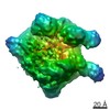
 Movie
Movie Controller


+ Open data

- Basic information
| Entry | Database: PDB / ID: 1im2 | ||||||
|---|---|---|---|---|---|---|---|
| Title | HslU, Haemophilus Influenzae, Selenomethionine Variant | ||||||
 Components Components | ATP-DEPENDENT HSL PROTEASE ATP-BINDING SUBUNIT HSLU | ||||||
 Keywords Keywords | CHAPERONE / AAA family | ||||||
| Function / homology |  Function and homology information Function and homology informationHslUV protease complex / proteasome-activating activity / protein unfolding / : / peptidase activity / ATP hydrolysis activity / ATP binding Similarity search - Function | ||||||
| Biological species |  Haemophilus influenzae (bacteria) Haemophilus influenzae (bacteria) | ||||||
| Method |  X-RAY DIFFRACTION / SYNCHROTRON / MOLECULAR REPLACEMENT / Resolution: 2.8 Å X-RAY DIFFRACTION / SYNCHROTRON / MOLECULAR REPLACEMENT / Resolution: 2.8 Å | ||||||
 Authors Authors | Trame, C.B. / McKay, D.B. | ||||||
 Citation Citation | Journal: Acta Crystallogr.,Sect.D / Year: 2001 Title: Structure of Haemophilus influenzae HslU protein in crystals with one-dimensional disorder twinning. Authors: Trame, C.B. / McKay, D.B. #1: Journal: Nature / Year: 2000Title: The Structures of HsIU and the ATP-dependent Protease HsIU-HsIV Authors: Bochtler, M. / Hartmann, C. / Song, H.K. / Bourenkov, G.P. / Bartunik, H.D. / Huber, R. #2: Journal: Proc.Natl.Acad.Sci.USA / Year: 2000Title: Mutational Studies on HslU and its Docking Mode with HslV Authors: Song, H.K. / Hartmann, C. / Ramachandran, R. / Bochtler, M. / Behrendt, R. / Moroder, L. / Huber, R. #3: Journal: Cell(Cambridge,Mass.) / Year: 2000Title: Crystal and Solution Structures of an HslUV Protease-Chaperone Complex Authors: Sousa, M.C. / Trame, C.B. / Tsuruta, H. / Wilbanks, S.M. / Reddy, V.S. / McKay, D.B. | ||||||
| History |
|
- Structure visualization
| Structure viewer | Molecule: MolmilJmol/JSmol |
|---|
- Downloads & links
-Download
| PDBx/mmCIF format | 1im2.cif.gz | 79 KB | Display | PDBx/mmCIF format |
|---|---|---|---|---|
| PDB format | pdb1im2.ent.gz | 59.3 KB | Display | PDB format |
| PDBx/mmJSON format | 1im2.json.gz | Tree view | PDBx/mmJSON format | |
| Others |  Other downloads Other downloads |
-Validation report
| Arichive directory | https://data.pdbj.org/pub/pdb/validation_reports/im/1im2ftp://data.pdbj.org/pub/pdb/validation_reports/im/1im2 | HTTPS FTP |
|---|
-Related structure data
| Related structure data | 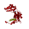 1g41SC S: Starting model for refinement C: citing same article ( |
|---|---|
| Similar structure data |


-Links
 PDBj
PDBj


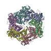
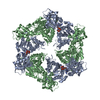
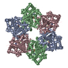
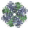
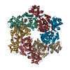
- Assembly
| Deposited unit | 
| ||||||||
|---|---|---|---|---|---|---|---|---|---|
| 1 | x 6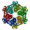
| ||||||||
| Unit cell |
| ||||||||
| Details | There are 2 molecules in the asymetric unit, because of twinning we observe only one (ref entry 1G41). The biological structure consists of hexamer which packs into dodecamer with 622 symmetry. |
-Components
| #1: Protein | Mass: 50144.922 Da / Num. of mol.: 1 Source method: isolated from a genetically manipulated source Source: (gene. exp.) Haemophilus influenzae (bacteria) / Plasmid: PRSET / Production host: |
|---|---|
| #2: Chemical | ChemComp-SO4 /   Mass: 96.063 Da / Num. of mol.: 1 / Source method: obtained synthetically / Formula: SO4 Mass: 96.063 Da / Num. of mol.: 1 / Source method: obtained synthetically / Formula: SO4 |
| #3: Chemical | ChemComp-ADP /   Mass: 427.201 Da / Num. of mol.: 1 / Source method: obtained synthetically / Formula: C10H15N5O10P2 / Comment: ADP, energy-carrying molecule*YM Mass: 427.201 Da / Num. of mol.: 1 / Source method: obtained synthetically / Formula: C10H15N5O10P2 / Comment: ADP, energy-carrying molecule*YM |
| Has protein modification | Y |
-Experimental details
-Experiment
| Experiment | Method: X-RAY DIFFRACTION / Number of used crystals: 1 |
|---|
- Sample preparation
| Crystal | Density Matthews: 5.45 Å3/Da | ||||||||||||||||||||||||||||||||||||||||||||||||||||||||||||||||||||||||||||||
|---|---|---|---|---|---|---|---|---|---|---|---|---|---|---|---|---|---|---|---|---|---|---|---|---|---|---|---|---|---|---|---|---|---|---|---|---|---|---|---|---|---|---|---|---|---|---|---|---|---|---|---|---|---|---|---|---|---|---|---|---|---|---|---|---|---|---|---|---|---|---|---|---|---|---|---|---|---|---|---|
| Crystal grow | Temperature: 291 K / Method: vapor diffusion, hanging drop / pH: 7.3 Details: PEG 2000 MME, lithium sulphate, magnesium sulphate, zink sulphate, tricine, europium chloride, ADP, pH 7.3, VAPOR DIFFUSION, HANGING DROP, temperature 291.0K | ||||||||||||||||||||||||||||||||||||||||||||||||||||||||||||||||||||||||||||||
| Crystal grow | *PLUS pH: 7 / Method: vapor diffusion | ||||||||||||||||||||||||||||||||||||||||||||||||||||||||||||||||||||||||||||||
| Components of the solutions | *PLUS
|
-Data collection
| Diffraction | Mean temperature: 100 K | ||||||||||||
|---|---|---|---|---|---|---|---|---|---|---|---|---|---|
| Diffraction source | Source: SYNCHROTRON / Site: SSRL  / Beamline: BL1-5 / Wavelength: 0.925, 0.979, 1.068 / Beamline: BL1-5 / Wavelength: 0.925, 0.979, 1.068 | ||||||||||||
| Detector | Type: ADSC QUANTUM 4 / Detector: CCD / Date: May 3, 1999 / Details: mirrors | ||||||||||||
| Radiation | Monochromator: SAGITALLY FOCUSED Si(111) / Protocol: MAD / Monochromatic (M) / Laue (L): M / Scattering type: x-ray | ||||||||||||
| Radiation wavelength |
| ||||||||||||
| Reflection | Resolution: 2.8→40 Å / Num. all: 47257 / Num. obs: 43125 / % possible obs: 90.5 % / Observed criterion σ(F): 2 / Observed criterion σ(I): 2 / Redundancy: 7 % / Biso Wilson estimate: 19.1 Å2 / Rmerge(I) obs: 0.112 / Rsym value: 0.112 / Net I/σ(I): 8.3 | ||||||||||||
| Reflection shell | Resolution: 2.8→2.85 Å / Redundancy: 3 % / Rmerge(I) obs: 0.305 / Mean I/σ(I) obs: 4.8 / Num. unique all: 1350 / Rsym value: 0.305 / % possible all: 61.3 | ||||||||||||
| Reflection | *PLUS Num. measured all: 190830 | ||||||||||||
| Reflection shell | *PLUS % possible obs: 61.3 % |
- Processing
| Software |
| |||||||||||||||||||||||||
|---|---|---|---|---|---|---|---|---|---|---|---|---|---|---|---|---|---|---|---|---|---|---|---|---|---|---|
| Refinement | Method to determine structure: MOLECULAR REPLACEMENT Starting model: PDB entry 1G41 Resolution: 2.8→37.9 Å / Rfactor Rfree error: 0.004 / Data cutoff high absF: 235868.52 / Data cutoff low absF: 0 / Isotropic thermal model: GROUP / Cross valid method: THROUGHOUT / σ(F): 2 / σ(I): 2 / Stereochemistry target values: Engh & Huber Details: TWINNING IN THE CRYSTAL PRODUCES THE P 622 SPACE GROUP AND AN EXTENDED UNIT CELL. REFINEMENT WAS PERFORMED IN THIS SETTING FOR ONE MOLECULE IN THE ASYMMETRIC UNIT. PLEASE SEE JOURNAL ...Details: TWINNING IN THE CRYSTAL PRODUCES THE P 622 SPACE GROUP AND AN EXTENDED UNIT CELL. REFINEMENT WAS PERFORMED IN THIS SETTING FOR ONE MOLECULE IN THE ASYMMETRIC UNIT. PLEASE SEE JOURNAL CITATION FOR ADDITIONAL DETAILS.
| |||||||||||||||||||||||||
| Solvent computation | Solvent model: FLAT MODEL / Bsol: 31.6 Å2 / ksol: 0.337 e/Å3 | |||||||||||||||||||||||||
| Displacement parameters | Biso mean: 57.6 Å2
| |||||||||||||||||||||||||
| Refine analyze |
| |||||||||||||||||||||||||
| Refinement step | Cycle: LAST / Resolution: 2.8→37.9 Å
| |||||||||||||||||||||||||
| Refine LS restraints |
| |||||||||||||||||||||||||
| LS refinement shell | Resolution: 2.8→2.98 Å / Rfactor Rfree error: 0.021 / Total num. of bins used: 6
| |||||||||||||||||||||||||
| Xplor file |
| |||||||||||||||||||||||||
| Software | *PLUS Name: CNS / Version: 1 / Classification: refinement | |||||||||||||||||||||||||
| Refinement | *PLUS σ(F): 2 / % reflection Rfree: 9.8 % / Rfactor Rfree: 0.25 | |||||||||||||||||||||||||
| Solvent computation | *PLUS | |||||||||||||||||||||||||
| Displacement parameters | *PLUS Biso mean: 57.6 Å2 | |||||||||||||||||||||||||
| Refine LS restraints | *PLUS
| |||||||||||||||||||||||||
| LS refinement shell | *PLUS Rfactor Rfree: 0.419 / % reflection Rfree: 9.9 % / Rfactor Rwork: 0.425 |
