 Movie
Movie Controller

[English] 日本語
 Yorodumi
- PDB-1do2: TRIGONAL CRYSTAL FORM OF HEAT SHOCK LOCUS U (HSLU) FROM ESCHERICH... -
+ Open data

- Basic information
| Entry | Database: PDB / ID: 1do2 | ||||||
|---|---|---|---|---|---|---|---|
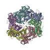
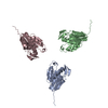
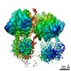
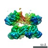
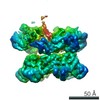
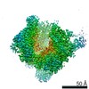
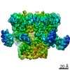
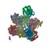
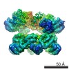
| Title | TRIGONAL CRYSTAL FORM OF HEAT SHOCK LOCUS U (HSLU) FROM ESCHERICHIA COLI | ||||||
 Components Components | PROTEIN (HEAT SHOCK LOCUS U) | ||||||
 Keywords Keywords | CHAPERONE / HSLU / CLPY / AAA-ATPASE / ATP-DEPENDENT PROTEOLYSIS / PROTEASOME | ||||||
| Function / homology |  Function and homology information Function and homology informationprotein denaturation / HslUV protease complex / proteasome-activating activity / protein unfolding / : / peptidase activity / cellular response to heat / response to heat / protein domain specific binding / magnesium ion binding ...protein denaturation / HslUV protease complex / proteasome-activating activity / protein unfolding / : / peptidase activity / cellular response to heat / response to heat / protein domain specific binding / magnesium ion binding / ATP hydrolysis activity / proteolysis / ATP binding / membrane / identical protein binding / cytosol Similarity search - Function | ||||||
| Biological species |  | ||||||
| Method |  X-RAY DIFFRACTION / SYNCHROTRON / MOLECULAR REPLACEMENT / Resolution: 4 Å X-RAY DIFFRACTION / SYNCHROTRON / MOLECULAR REPLACEMENT / Resolution: 4 Å | ||||||
 Authors Authors | Bochtler, M. / Hartmann, C. / Song, H.K. / Bourenkov, G.P. / Bartunik, H.D. | ||||||
 Citation Citation | Journal: Nature / Year: 2000 Title: The structures of HsIU and the ATP-dependent protease HsIU-HsIV. Authors: Bochtler, M. / Hartmann, C. / Song, H.K. / Bourenkov, G.P. / Bartunik, H.D. / Huber, R. #1: Journal: Proc.Natl.Acad.Sci.USA / Year: 1997Title: Crystal Structure of Heat Shock Locus V (HslV) from Escherichia coli Authors: Bochtler, M. / Ditzel, L. / Groll, M. / Huber, R. #2: Journal: Proc.Natl.Acad.Sci.USA / Year: 1996Title: HslV-HslU: A Novel ATP-Dependent Protease Complex in Escherichia coli Related to the Eukaryotic Proteasome Authors: Rohrwild, M. / Coux, O. / Huang, H.C. / Moerschell, R.P. / Yoo, S.J. / Seol, J.H. / Chung, C.H. / Goldberg, A.L. #3: Journal: Gene / Year: 1993Title: Sequence Analysis of Four New Heat-Shock Genes Constituting the hslTS/ibpAB and hslVU Operons in Escherichia coli Authors: Chuang, S.E. / Burland, V. / Plunket III, G. / Daniels, D.L. / Blattner, F.R. | ||||||
| History |
|
- Structure visualization
| Structure viewer | Molecule: MolmilJmol/JSmol |
|---|
- Downloads & links
-Download
| PDBx/mmCIF format | 1do2.cif.gz | 325.3 KB | Display | PDBx/mmCIF format |
|---|---|---|---|---|
| PDB format | pdb1do2.ent.gz | 261.4 KB | Display | PDB format |
| PDBx/mmJSON format | 1do2.json.gz | Tree view | PDBx/mmJSON format | |
| Others |  Other downloads Other downloads |
-Validation report
| Arichive directory | https://data.pdbj.org/pub/pdb/validation_reports/do/1do2ftp://data.pdbj.org/pub/pdb/validation_reports/do/1do2 | HTTPS FTP |
|---|
-Related structure data

-Links
 PDBj
PDBj


- Assembly
| Deposited unit | 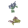
| ||||||||
|---|---|---|---|---|---|---|---|---|---|
| 1 | 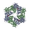
| ||||||||
| 2 | 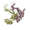
| ||||||||
| 3 | 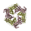
| ||||||||
| Unit cell |
|
-Components
| #1: Protein | Mass: 49528.508 Da / Num. of mol.: 4 Source method: isolated from a genetically manipulated source Source: (gene. exp.) #2: Chemical |   Mass: 506.196 Da / Num. of mol.: 2 / Source method: obtained synthetically / Formula: C10H17N6O12P3 / Comment: AMP-PNP, energy-carrying molecule analogue*YM Mass: 506.196 Da / Num. of mol.: 2 / Source method: obtained synthetically / Formula: C10H17N6O12P3 / Comment: AMP-PNP, energy-carrying molecule analogue*YMSequence details | EDMAN-DEGRADATION HAS SHOWN THAT THE AMINOTERMINAL METHIONINE IS CLEAVED IN THE WILD TYPE. ...EDMAN-DEGRADATIO | |
|---|
-Experimental details
-Experiment
| Experiment | Method: X-RAY DIFFRACTION / Number of used crystals: 1 |
|---|
- Sample preparation
| Crystal | Density Matthews: 5.09 Å3/Da / Density % sol: 75.84 % | |||||||||||||||||||||||||||||||||||||||||||||||||||||||||||||||
|---|---|---|---|---|---|---|---|---|---|---|---|---|---|---|---|---|---|---|---|---|---|---|---|---|---|---|---|---|---|---|---|---|---|---|---|---|---|---|---|---|---|---|---|---|---|---|---|---|---|---|---|---|---|---|---|---|---|---|---|---|---|---|---|---|
| Crystal grow | Temperature: 291 K / Method: vapor diffusion, sitting drop / pH: 6.5 Details: reservoir: 100 mM sodium cacodylate, 15% glycerol, 10.5% PEG8000, 500 mM (NH4)2(SO4). drop: 0.002 ml reservoir solution, 0.002 ml 16 mg/ml protein in 20 mM Tris/HCl, pH 7.5, 5 mM MgCl2, 1 mM ...Details: reservoir: 100 mM sodium cacodylate, 15% glycerol, 10.5% PEG8000, 500 mM (NH4)2(SO4). drop: 0.002 ml reservoir solution, 0.002 ml 16 mg/ml protein in 20 mM Tris/HCl, pH 7.5, 5 mM MgCl2, 1 mM AMP-PNP, 1 mM NaN3, 0.0005 ml 1.0 M guanidinium chloride, 0.001 ml C12E8 , pH 6.5, VAPOR DIFFUSION, SITTING DROP, temperature 291K | |||||||||||||||||||||||||||||||||||||||||||||||||||||||||||||||
| Crystal grow | *PLUS Details: drop consists of equal volume of protein and reservoir solutions in Buffer B | |||||||||||||||||||||||||||||||||||||||||||||||||||||||||||||||
| Components of the solutions | *PLUS
|
-Data collection
| Diffraction | Mean temperature: 100 K |
|---|---|
| Diffraction source | Source: SYNCHROTRON / Site: MPG/DESY, HAMBURG  / Beamline: BW6 / Wavelength: 1.0712 / Beamline: BW6 / Wavelength: 1.0712 |
| Detector | Type: MARRESEARCH / Detector: CCD / Date: Jun 15, 1999 / Details: MIRROR |
| Radiation | Monochromator: DOUBLE CRYSTAL / Protocol: SINGLE WAVELENGTH / Monochromatic (M) / Laue (L): M / Scattering type: x-ray |
| Radiation wavelength | Wavelength: 1.0712 Å / Relative weight: 1 |
| Reflection | Resolution: 4→20 Å / Num. all: 224179 / Num. obs: 32750 / % possible obs: 95.8 % / Redundancy: 6 % / Biso Wilson estimate: 50.4 Å2 / Rmerge(I) obs: 0.105 / Rsym value: 10.5 |
| Reflection shell | Resolution: 4→4.07 Å / Redundancy: 3.2 % / Rmerge(I) obs: 0.337 / Rsym value: 33.7 / % possible all: 96.5 |
| Reflection | *PLUS Num. measured all: 224179 |
| Reflection shell | *PLUS % possible obs: 96.5 % |
- Processing
| Software |
| ||||||||||||||||||||||||||||||||||||||||||||||||||||||||||||||||||||||||||||||||
|---|---|---|---|---|---|---|---|---|---|---|---|---|---|---|---|---|---|---|---|---|---|---|---|---|---|---|---|---|---|---|---|---|---|---|---|---|---|---|---|---|---|---|---|---|---|---|---|---|---|---|---|---|---|---|---|---|---|---|---|---|---|---|---|---|---|---|---|---|---|---|---|---|---|---|---|---|---|---|---|---|---|
| Refinement | Method to determine structure: MOLECULAR REPLACEMENT Starting model: HSLU FROM HSLU IN HSLVU CRYSTALS Resolution: 4→15 Å / Rfactor Rfree error: 0.007 / Data cutoff low absF: 0 / Isotropic thermal model: RESTRAINED / Cross valid method: THROUGHOUT / σ(F): 0
| ||||||||||||||||||||||||||||||||||||||||||||||||||||||||||||||||||||||||||||||||
| Displacement parameters | Biso mean: 50.4 Å2
| ||||||||||||||||||||||||||||||||||||||||||||||||||||||||||||||||||||||||||||||||
| Refine analyze |
| ||||||||||||||||||||||||||||||||||||||||||||||||||||||||||||||||||||||||||||||||
| Refinement step | Cycle: LAST / Resolution: 4→15 Å
| ||||||||||||||||||||||||||||||||||||||||||||||||||||||||||||||||||||||||||||||||
| Refine LS restraints |
| ||||||||||||||||||||||||||||||||||||||||||||||||||||||||||||||||||||||||||||||||
| LS refinement shell | Resolution: 4→4.25 Å / Rfactor Rfree error: 0.02 / Total num. of bins used: 6
| ||||||||||||||||||||||||||||||||||||||||||||||||||||||||||||||||||||||||||||||||
| Xplor file |
|
