 Movie
Movie Controller

[English] 日本語
 Yorodumi
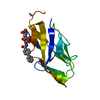
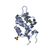
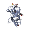
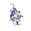
- PDB-1od7: N-terminal of Sialoadhesin in complex with Me-a-9-N-(naphthyl-2-c... -
+ Open data

- Basic information
| Entry | Database: PDB / ID: 1od7 | ||||||
|---|---|---|---|---|---|---|---|
| Title | N-terminal of Sialoadhesin in complex with Me-a-9-N-(naphthyl-2-carbonyl)-amino-9-deoxy-Neu5Ac (NAP compound) | ||||||
 Components Components | SIALOADHESIN | ||||||
 Keywords Keywords | LECTIN/IMMNUE SYSTEM / IMMUNE SYSTEM / IMMUNOGLOBULIN SUPERFAMILY / CARBOHYDRATE BINDING / SIGLEC / INHIBITOR DESIGN / CELL ADHESION / LECTIN-IMMNUE SYSTEM complex | ||||||
| Function / homology |  Function and homology information Function and homology informationpositive regulation of T cell apoptotic process / virion binding / Immunoregulatory interactions between a Lymphoid and a non-Lymphoid cell / positive regulation of extrinsic apoptotic signaling pathway / negative regulation of type I interferon production / positive regulation of type I interferon production / late endosome / carbohydrate binding / clathrin-dependent endocytosis of virus by host cell / early endosome ...positive regulation of T cell apoptotic process / virion binding / Immunoregulatory interactions between a Lymphoid and a non-Lymphoid cell / positive regulation of extrinsic apoptotic signaling pathway / negative regulation of type I interferon production / positive regulation of type I interferon production / late endosome / carbohydrate binding / clathrin-dependent endocytosis of virus by host cell / early endosome / cell adhesion / extracellular region / plasma membrane Similarity search - Function | ||||||
| Biological species |  | ||||||
| Method |  X-RAY DIFFRACTION / SYNCHROTRON / MOLECULAR REPLACEMENT / Resolution: 3 Å X-RAY DIFFRACTION / SYNCHROTRON / MOLECULAR REPLACEMENT / Resolution: 3 Å | ||||||
 Authors Authors | Zaccai, N.R. / Maenaka, K. / Maenaka, T. / Crocker, P.R. / Brossmer, R. / Kelm, S. / Jones, E.Y. | ||||||
 Citation Citation | Journal: Structure / Year: 2003 Title: Structure-Guided Design of Sialic Acid-Based Siglec Inhibitors and Crystallographic Analysis in Complex with Sialoadhesin Authors: Zaccai, N.R. / Maenaka, K. / Maenaka, T. / Crocker, P.R. / Brossmer, R. / Kelm, S. / Jones, E.Y. | ||||||
| History |
| ||||||
| Remark 700 | SHEET THE SHEET STRUCTURE OF THIS MOLECULE IS BIFURCATED. IN ORDER TO REPRESENT THIS FEATURE IN ... SHEET THE SHEET STRUCTURE OF THIS MOLECULE IS BIFURCATED. IN ORDER TO REPRESENT THIS FEATURE IN THE SHEET RECORDS BELOW, TWO SHEETS ARE DEFINED. |
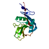
- Structure visualization
| Structure viewer | Molecule: MolmilJmol/JSmol |
|---|
- Downloads & links
-Download
| PDBx/mmCIF format | 1od7.cif.gz | 36.8 KB | Display | PDBx/mmCIF format |
|---|---|---|---|---|
| PDB format | pdb1od7.ent.gz | 24.9 KB | Display | PDB format |
| PDBx/mmJSON format | 1od7.json.gz | Tree view | PDBx/mmJSON format | |
| Others |  Other downloads Other downloads |
-Validation report
| Arichive directory | https://data.pdbj.org/pub/pdb/validation_reports/od/1od7ftp://data.pdbj.org/pub/pdb/validation_reports/od/1od7 | HTTPS FTP |
|---|
-Related structure data
| Related structure data |  1od9C  1odaC  1qfoS S: Starting model for refinement C: citing same article ( |
|---|---|
| Similar structure data |


-Links
 PDBj
PDBj


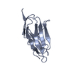
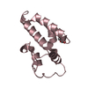
- Assembly
| Deposited unit | 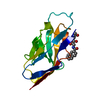
| ||||||||
|---|---|---|---|---|---|---|---|---|---|
| 1 |
| ||||||||
| Unit cell |
|
-Components
| #1: Protein | Mass: 13319.107 Da / Num. of mol.: 1 Fragment: DOMAIN ONE, IG-LIKE V-TYPE DOMAIN, RESIDUES 20-138 Source method: isolated from a genetically manipulated source Details: BOUND TO ME-A-9-N-(NAPHTHYL-2-CARBONYL)-AMINO-9-DEOXY-NEU5AC Source: (gene. exp.)  CRICETULUS GRISEUS (Chinese hamster) / References: UniProt: Q62230 CRICETULUS GRISEUS (Chinese hamster) / References: UniProt: Q62230 |
|---|---|
| #2: Chemical | ChemComp-SUW / 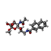  Mass: 476.476 Da / Num. of mol.: 1 / Source method: obtained synthetically / Formula: C23H28N2O9 Mass: 476.476 Da / Num. of mol.: 1 / Source method: obtained synthetically / Formula: C23H28N2O9 |
| Compound details | THIS IS A MACROPHAGE-RESTRICTED ADHESION MOLECULE THAT MEDIATES SIALIC-ACID DEPENDENT BINDING TO ...THIS IS A MACROPHAGE |
| Has protein modification | Y |
-Experimental details
-Experiment
| Experiment | Method: X-RAY DIFFRACTION / Number of used crystals: 1 |
|---|
- Sample preparation
| Crystal | Density Matthews: 2.54 Å3/Da / Density % sol: 52 % | ||||||||||||||||||||||||||||||||||||||||||||||||
|---|---|---|---|---|---|---|---|---|---|---|---|---|---|---|---|---|---|---|---|---|---|---|---|---|---|---|---|---|---|---|---|---|---|---|---|---|---|---|---|---|---|---|---|---|---|---|---|---|---|
| Crystal grow | pH: 5.6 Details: 30 % (W/V) PEG 4000, 10 MM DTT, 0.1M SODIUM HEPES PH 5.6,25MM NAP COMPOUND | ||||||||||||||||||||||||||||||||||||||||||||||||
| Crystal grow | *PLUS Temperature: 17 ℃ / Method: vapor diffusion, sitting drop | ||||||||||||||||||||||||||||||||||||||||||||||||
| Components of the solutions | *PLUS
|
-Data collection
| Diffraction | Mean temperature: 110 K |
|---|---|
| Diffraction source | Source: SYNCHROTRON / Site: SRS  / Beamline: PX14.2 / Wavelength: 0.97 / Beamline: PX14.2 / Wavelength: 0.97 |
| Detector | Type: MARRESEARCH / Detector: CCD |
| Radiation | Protocol: SINGLE WAVELENGTH / Monochromatic (M) / Laue (L): M / Scattering type: x-ray |
| Radiation wavelength | Wavelength: 0.97 Å / Relative weight: 1 |
| Reflection | Resolution: 3→25 Å / Num. obs: 3047 / % possible obs: 97 % / Observed criterion σ(I): 0 / Redundancy: 9.6 % / Biso Wilson estimate: 26.1 Å2 / Rmerge(I) obs: 0.24 / Net I/σ(I): 8.4 |
| Reflection shell | Resolution: 3→3.11 Å / Rmerge(I) obs: 0.51 / Mean I/σ(I) obs: 2.1 / % possible all: 72.5 |
| Reflection | *PLUS Highest resolution: 3 Å / Lowest resolution: 25 Å / % possible obs: 97 % / Num. measured all: 29346 / Rmerge(I) obs: 0.24 |
| Reflection shell | *PLUS Highest resolution: 3 Å / % possible obs: 72.5 % / Rmerge(I) obs: 0.506 / Mean I/σ(I) obs: 2.1 |
- Processing
| Software |
| ||||||||||||||||||||||||||||||||||||||||||||||||||||||||||||||||||||||||||||||||
|---|---|---|---|---|---|---|---|---|---|---|---|---|---|---|---|---|---|---|---|---|---|---|---|---|---|---|---|---|---|---|---|---|---|---|---|---|---|---|---|---|---|---|---|---|---|---|---|---|---|---|---|---|---|---|---|---|---|---|---|---|---|---|---|---|---|---|---|---|---|---|---|---|---|---|---|---|---|---|---|---|---|
| Refinement | Method to determine structure: MOLECULAR REPLACEMENT Starting model: DOMAIN A OF PDB ENTRY 1QFO Resolution: 3→19.54 Å / Rfactor Rfree error: 0.023 / Data cutoff high absF: 118124.86 / Data cutoff low absF: 0 / Isotropic thermal model: RESTRAINED / Cross valid method: THROUGHOUT / σ(F): 0 / Details: BULK SOLVENT MODEL USED
| ||||||||||||||||||||||||||||||||||||||||||||||||||||||||||||||||||||||||||||||||
| Solvent computation | Solvent model: FLAT MODEL / Bsol: 10 Å2 / ksol: 0.32 e/Å3 | ||||||||||||||||||||||||||||||||||||||||||||||||||||||||||||||||||||||||||||||||
| Displacement parameters | Biso mean: 21.6 Å2
| ||||||||||||||||||||||||||||||||||||||||||||||||||||||||||||||||||||||||||||||||
| Refine analyze |
| ||||||||||||||||||||||||||||||||||||||||||||||||||||||||||||||||||||||||||||||||
| Refinement step | Cycle: LAST / Resolution: 3→19.54 Å
| ||||||||||||||||||||||||||||||||||||||||||||||||||||||||||||||||||||||||||||||||
| Refine LS restraints |
| ||||||||||||||||||||||||||||||||||||||||||||||||||||||||||||||||||||||||||||||||
| LS refinement shell | Resolution: 3→3.19 Å / Rfactor Rfree error: 0.082 / Total num. of bins used: 6
| ||||||||||||||||||||||||||||||||||||||||||||||||||||||||||||||||||||||||||||||||
| Xplor file |
| ||||||||||||||||||||||||||||||||||||||||||||||||||||||||||||||||||||||||||||||||
| Refinement | *PLUS Highest resolution: 3 Å | ||||||||||||||||||||||||||||||||||||||||||||||||||||||||||||||||||||||||||||||||
| Solvent computation | *PLUS | ||||||||||||||||||||||||||||||||||||||||||||||||||||||||||||||||||||||||||||||||
| Displacement parameters | *PLUS | ||||||||||||||||||||||||||||||||||||||||||||||||||||||||||||||||||||||||||||||||
| Refine LS restraints | *PLUS
|
