 Movie
Movie Controller

[English] 日本語
 Yorodumi
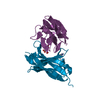
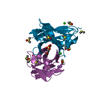
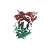
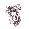
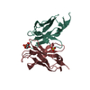
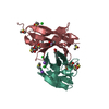
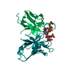
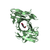
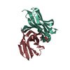
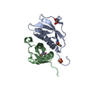
- PDB-1oax: Fv Structure of the IgE SPE-7 in complex with acenaphthenequinone -
+ Open data

- Basic information
| Entry | Database: PDB / ID: 1oax | ||||||
|---|---|---|---|---|---|---|---|
| Title | Fv Structure of the IgE SPE-7 in complex with acenaphthenequinone | ||||||
 Components Components | (IMMUNOGLOBULIN E) x 2 | ||||||
 Keywords Keywords | IMMUNE SYSTEM / ANTIBODY-COMPLEX / ANTIBODY / ALLERGY / IGE | ||||||
| Function / homology |  Function and homology information Function and homology information | ||||||
| Biological species |  | ||||||
| Method |  X-RAY DIFFRACTION / MOLECULAR REPLACEMENT / Resolution: 2.67 Å X-RAY DIFFRACTION / MOLECULAR REPLACEMENT / Resolution: 2.67 Å | ||||||
 Authors Authors | James, L.C. / Roversi, P. / Tawfik, D. | ||||||
 Citation Citation | Journal: Science / Year: 2003 Title: Antibody Multispecificity Mediated by Conformational Diversity Authors: James, L.C. / Roversi, P. / Tawfik, D. | ||||||
| History |
| ||||||
| Remark 700 | SHEET THE SHEET STRUCTURE OF THIS MOLECULE IS BIFURCATED. IN ORDER TO REPRESENT THIS FEATURE IN ... SHEET THE SHEET STRUCTURE OF THIS MOLECULE IS BIFURCATED. IN ORDER TO REPRESENT THIS FEATURE IN THE SHEET RECORDS BELOW, TWO SHEETS ARE DEFINED. |
- Structure visualization
| Structure viewer | Molecule: MolmilJmol/JSmol |
|---|
- Downloads & links
-Download
| PDBx/mmCIF format | 1oax.cif.gz | 138.7 KB | Display | PDBx/mmCIF format |
|---|---|---|---|---|
| PDB format | pdb1oax.ent.gz | 110.6 KB | Display | PDB format |
| PDBx/mmJSON format | 1oax.json.gz | Tree view | PDBx/mmJSON format | |
| Others |  Other downloads Other downloads |
-Validation report
| Arichive directory | https://data.pdbj.org/pub/pdb/validation_reports/oa/1oaxftp://data.pdbj.org/pub/pdb/validation_reports/oa/1oax | HTTPS FTP |
|---|
-Related structure data
| Related structure data |  1oaqC  1oarC  1oauC  1oayC  1oazC  1ocwC  1anqS C: citing same article ( S: Starting model for refinement |
|---|---|
| Similar structure data |
-Links
 PDBj
PDBj

- Assembly
| Deposited unit | 
| |||||||||||||||||||||||||||||||||||||||||||||||||||||||||||||||||||||||||||||||||||||||||||
|---|---|---|---|---|---|---|---|---|---|---|---|---|---|---|---|---|---|---|---|---|---|---|---|---|---|---|---|---|---|---|---|---|---|---|---|---|---|---|---|---|---|---|---|---|---|---|---|---|---|---|---|---|---|---|---|---|---|---|---|---|---|---|---|---|---|---|---|---|---|---|---|---|---|---|---|---|---|---|---|---|---|---|---|---|---|---|---|---|---|---|---|---|
| 1 | 
| |||||||||||||||||||||||||||||||||||||||||||||||||||||||||||||||||||||||||||||||||||||||||||
| 2 | 
| |||||||||||||||||||||||||||||||||||||||||||||||||||||||||||||||||||||||||||||||||||||||||||
| 3 | 
| |||||||||||||||||||||||||||||||||||||||||||||||||||||||||||||||||||||||||||||||||||||||||||
| 4 | 
| |||||||||||||||||||||||||||||||||||||||||||||||||||||||||||||||||||||||||||||||||||||||||||
| Unit cell |
| |||||||||||||||||||||||||||||||||||||||||||||||||||||||||||||||||||||||||||||||||||||||||||
| Noncrystallographic symmetry (NCS) | NCS domain:
NCS domain segments:
NCS ensembles :
NCS oper:
|
-Components
| #1: Antibody | Mass: 13671.359 Da / Num. of mol.: 2 / Fragment: FV REGION, RESIDUES 1-122 Source method: isolated from a genetically manipulated source Source: (gene. exp.)  #2: Antibody | Mass: 11572.913 Da / Num. of mol.: 4 / Fragment: FV REGION, RESIDUES 1-110 Source method: isolated from a genetically manipulated source Source: (gene. exp.) #3: Chemical | 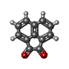  Mass: 182.175 Da / Num. of mol.: 2 / Source method: obtained synthetically / Formula: C12H6O2 Mass: 182.175 Da / Num. of mol.: 2 / Source method: obtained synthetically / Formula: C12H6O2#4: Water | ChemComp-HOH / |  Mass: 18.015 Da / Num. of mol.: 70 / Source method: isolated from a natural source / Formula: H2O Mass: 18.015 Da / Num. of mol.: 70 / Source method: isolated from a natural source / Formula: H2OHas protein modification | Y | |
|---|
-Experimental details
-Experiment
| Experiment | Method: X-RAY DIFFRACTION / Number of used crystals: 1 |
|---|
- Sample preparation
| Crystal | Density Matthews: 1.5 Å3/Da / Density % sol: 70 % |
|---|---|
| Crystal grow | pH: 5.5 Details: 21% PEG 8K 0.1M NA CACODYLATE, 0.2M NA ACETATE PH5.5, pH 5.50 |
-Data collection
| Diffraction | Mean temperature: 100 K |
|---|---|
| Diffraction source | Source: ROTATING ANODE / Type: RIGAKU RU200 / Wavelength: 1.5418 |
| Detector | Type: MARRESEARCH / Detector: IMAGE PLATE / Date: Oct 15, 2001 / Details: MIRRORS |
| Radiation | Monochromator: NI FILTER / Protocol: SINGLE WAVELENGTH / Monochromatic (M) / Laue (L): M / Scattering type: x-ray |
| Radiation wavelength | Wavelength: 1.5418 Å / Relative weight: 1 |
| Reflection | Resolution: 2.66→39.5 Å / Num. obs: 30262 / % possible obs: 97.4 % / Observed criterion σ(I): 2 / Redundancy: 9.9 % / Rsym value: 0.051 / Net I/σ(I): 9.3 |
| Reflection shell | Resolution: 2.67→2.98 Å / Redundancy: 6.7 % / Rmerge(I) obs: 0.234 / Mean I/σ(I) obs: 3.6 / % possible all: 91.7 |
- Processing
| Software |
| ||||||||||||||||||||||||||||||||||||||||||||||||||||||||||||||||||||||||||||||||||||||||||||||||||||||||||||||||||||||||||||||||||||||||||||||||||||||||||||||||||||||||||||||||||||||
|---|---|---|---|---|---|---|---|---|---|---|---|---|---|---|---|---|---|---|---|---|---|---|---|---|---|---|---|---|---|---|---|---|---|---|---|---|---|---|---|---|---|---|---|---|---|---|---|---|---|---|---|---|---|---|---|---|---|---|---|---|---|---|---|---|---|---|---|---|---|---|---|---|---|---|---|---|---|---|---|---|---|---|---|---|---|---|---|---|---|---|---|---|---|---|---|---|---|---|---|---|---|---|---|---|---|---|---|---|---|---|---|---|---|---|---|---|---|---|---|---|---|---|---|---|---|---|---|---|---|---|---|---|---|---|---|---|---|---|---|---|---|---|---|---|---|---|---|---|---|---|---|---|---|---|---|---|---|---|---|---|---|---|---|---|---|---|---|---|---|---|---|---|---|---|---|---|---|---|---|---|---|---|---|
| Refinement | Method to determine structure: MOLECULAR REPLACEMENT Starting model: PDB ENTRY 1ANQ Resolution: 2.67→39.53 Å / Cor.coef. Fo:Fc: 0.893 / Cor.coef. Fo:Fc free: 0.863 / SU B: 11.63 / SU ML: 0.248 / Cross valid method: THROUGHOUT / ESU R Free: 0.332 / Stereochemistry target values: MAXIMUM LIKELIHOOD Details: HYDROGENS HAVE BEEN ADDED IN THE RIDING POSITIONS. THIS ENTRY HAS 4 SETS OF 2 CHAINS WHICH ARE RELATED BY NCS. CHAINS I AND K IN THIS ENTRY ARE MOSTLY DISORDERED AND HENCE NO STRUCTURE WAS ...Details: HYDROGENS HAVE BEEN ADDED IN THE RIDING POSITIONS. THIS ENTRY HAS 4 SETS OF 2 CHAINS WHICH ARE RELATED BY NCS. CHAINS I AND K IN THIS ENTRY ARE MOSTLY DISORDERED AND HENCE NO STRUCTURE WAS CLEARLY DEFINED FOR THESE CHAINS.
| ||||||||||||||||||||||||||||||||||||||||||||||||||||||||||||||||||||||||||||||||||||||||||||||||||||||||||||||||||||||||||||||||||||||||||||||||||||||||||||||||||||||||||||||||||||||
| Solvent computation | Ion probe radii: 0.8 Å / Shrinkage radii: 0.8 Å / VDW probe radii: 1.4 Å / Solvent model: BABINET MODEL PLUS MASK | ||||||||||||||||||||||||||||||||||||||||||||||||||||||||||||||||||||||||||||||||||||||||||||||||||||||||||||||||||||||||||||||||||||||||||||||||||||||||||||||||||||||||||||||||||||||
| Displacement parameters | Biso mean: 40.95 Å2
| ||||||||||||||||||||||||||||||||||||||||||||||||||||||||||||||||||||||||||||||||||||||||||||||||||||||||||||||||||||||||||||||||||||||||||||||||||||||||||||||||||||||||||||||||||||||
| Refinement step | Cycle: LAST / Resolution: 2.67→39.53 Å
| ||||||||||||||||||||||||||||||||||||||||||||||||||||||||||||||||||||||||||||||||||||||||||||||||||||||||||||||||||||||||||||||||||||||||||||||||||||||||||||||||||||||||||||||||||||||
| Refine LS restraints |
|