 Movie
Movie Controller


+ Open data

- Basic information
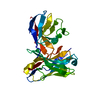
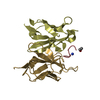
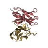
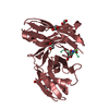
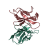
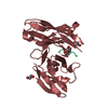
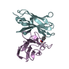
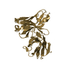
| Entry | Database: PDB / ID: 1oaq | ||||||
|---|---|---|---|---|---|---|---|
| Title | Free conformation Ab1 of the IgE SPE-7 | ||||||
 Components Components |
| ||||||
 Keywords Keywords | IMMUNE SYSTEM / ALLERGY / IGE / CONFORMATIONAL DIVERSITY / MULTISPECIFICITY | ||||||
| Function / homology |  Function and homology information Function and homology information | ||||||
| Biological species |  | ||||||
| Method |  X-RAY DIFFRACTION / MOLECULAR REPLACEMENT / Resolution: 1.5 Å X-RAY DIFFRACTION / MOLECULAR REPLACEMENT / Resolution: 1.5 Å | ||||||
 Authors Authors | James, L.C. / Roversi, P. / Tawfik, D. | ||||||
 Citation Citation | Journal: Science / Year: 2003 Title: Antibody Multispecificity Mediated by Conformational Diversity Authors: James, L.C. / Roversi, P. / Tawfik, D. | ||||||
| History |
| ||||||
| Remark 700 | SHEET THE SHEET STRUCTURE OF THIS MOLECULE IS BIFURCATED. IN ORDER TO REPRESENT THIS FEATURE IN ... SHEET THE SHEET STRUCTURE OF THIS MOLECULE IS BIFURCATED. IN ORDER TO REPRESENT THIS FEATURE IN THE SHEET RECORDS BELOW, TWO SHEETS ARE DEFINED. |
- Structure visualization
| Structure viewer | Molecule: MolmilJmol/JSmol |
|---|
- Downloads & links
-Download
| PDBx/mmCIF format | 1oaq.cif.gz | 61.8 KB | Display | PDBx/mmCIF format |
|---|---|---|---|---|
| PDB format | pdb1oaq.ent.gz | 44.8 KB | Display | PDB format |
| PDBx/mmJSON format | 1oaq.json.gz | Tree view | PDBx/mmJSON format | |
| Others |  Other downloads Other downloads |
-Validation report
| Arichive directory | https://data.pdbj.org/pub/pdb/validation_reports/oa/1oaqftp://data.pdbj.org/pub/pdb/validation_reports/oa/1oaq | HTTPS FTP |
|---|
-Related structure data
| Related structure data |  1oarC  1oauC  1oaxC  1oayC  1oazC  1ocwC  1anqS C: citing same article ( S: Starting model for refinement |
|---|---|
| Similar structure data |


-Links
 PDBj
PDBj

- Assembly
| Deposited unit | 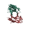
| ||||||||
|---|---|---|---|---|---|---|---|---|---|
| 1 |
| ||||||||
| Unit cell |
|
-Components
| #1: Antibody | Mass: 13616.237 Da / Num. of mol.: 1 / Fragment: FV, RESIDUES 1-121 Source method: isolated from a genetically manipulated source Source: (gene. exp.)  |
|---|---|
| #2: Antibody | Mass: 12512.920 Da / Num. of mol.: 1 / Fragment: FV, RESIDUES 1-120 Source method: isolated from a genetically manipulated source Source: (gene. exp.) |
| #3: Water | ChemComp-HOH /  Mass: 18.015 Da / Num. of mol.: 275 / Source method: isolated from a natural source / Formula: H2O Mass: 18.015 Da / Num. of mol.: 275 / Source method: isolated from a natural source / Formula: H2O |
| Has protein modification | Y |
| Sequence details | NEW SEQUENCE |
-Experimental details
-Experiment
| Experiment | Method: X-RAY DIFFRACTION / Number of used crystals: 1 |
|---|
- Sample preparation
| Crystal | Density Matthews: 1.5 Å3/Da / Density % sol: 70 % |
|---|---|
| Crystal grow | pH: 5 Details: 1.2M NA CITRATE 0.2M NA CACODYLATE PH5, 0.2M LITHIUM, pH 5.00 |
| Crystal grow | *PLUS pH: 6.2 / Method: vapor diffusion, hanging drop |
| Components of the solutions | *PLUS Conc.: 1 M / Common name: sodium citrate / Details: pH6.2 |
-Data collection
| Diffraction | Mean temperature: 100 K |
|---|---|
| Diffraction source | Source: ROTATING ANODE / Type: RIGAKU RU200 / Wavelength: 1.5418 |
| Detector | Type: MARRESEARCH / Detector: IMAGE PLATE / Date: Nov 15, 2000 / Details: MIRRORS |
| Radiation | Monochromator: NI FILTER / Protocol: SINGLE WAVELENGTH / Monochromatic (M) / Laue (L): M / Scattering type: x-ray |
| Radiation wavelength | Wavelength: 1.5418 Å / Relative weight: 1 |
| Reflection | Resolution: 1.5→33.3 Å / Num. obs: 34286 / % possible obs: 97.4 % / Observed criterion σ(I): 2 / Redundancy: 9.9 % / Rmerge(I) obs: 0.051 / Net I/σ(I): 9.3 |
| Reflection shell | Resolution: 1.5→1.58 Å / Redundancy: 6.7 % / Rmerge(I) obs: 0.234 / Mean I/σ(I) obs: 3.6 / % possible all: 91.7 |
| Reflection | *PLUS Highest resolution: 1.5 Å / Num. obs: 34284 / % possible obs: 92.8 % / Redundancy: 12.6 % / Rmerge(I) obs: 0.0431 |
| Reflection shell | *PLUS Highest resolution: 1.5 Å / % possible obs: 92.8 % / Rmerge(I) obs: 0.238 / Mean I/σ(I) obs: 1.2 |
- Processing
| Software |
| ||||||||||||||||||||
|---|---|---|---|---|---|---|---|---|---|---|---|---|---|---|---|---|---|---|---|---|---|
| Refinement | Method to determine structure: MOLECULAR REPLACEMENT Starting model: PDB ENTRY 1ANQ Resolution: 1.5→33.33 Å / SU B: 1.136 / SU ML: 0.043 / Cross valid method: THROUGHOUT / ESU R: 0.071 / ESU R Free: 0.07
| ||||||||||||||||||||
| Displacement parameters | Biso mean: 18.638 Å2
| ||||||||||||||||||||
| Refinement step | Cycle: LAST / Resolution: 1.5→33.33 Å
| ||||||||||||||||||||
| Refinement | *PLUS Highest resolution: 1.5 Å / Rfactor Rfree: 0.182 / Rfactor Rwork: 0.16 | ||||||||||||||||||||
| Solvent computation | *PLUS | ||||||||||||||||||||
| Displacement parameters | *PLUS | ||||||||||||||||||||
| Refine LS restraints | *PLUS
| ||||||||||||||||||||
| LS refinement shell | *PLUS Rfactor Rfree: 0.175 / Rfactor Rwork: 0.155 |
