 Movie
Movie Controller

[English] 日本語
 Yorodumi
- PDB-1dsf: THE CRYSTAL STRUCTURE OF THE DISULFIDE-STABILIZED FV FRAGMENT OF ... -
+ Open data

- Basic information
| Entry | Database: PDB / ID: 1dsf | ||||||
|---|---|---|---|---|---|---|---|
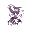
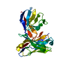
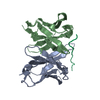
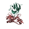
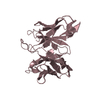
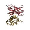
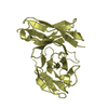
| Title | THE CRYSTAL STRUCTURE OF THE DISULFIDE-STABILIZED FV FRAGMENT OF ANTICANCER ANTIBODY B1: CONFORMATIONAL INFLUENCE OF AN ENGINEERED DISULFIDE BOND | ||||||
 Components Components | (ANTICANCER ANTIBODY B1) x 2 | ||||||
 Keywords Keywords | IMMUNOGLOBULIN / MONOCLONAL ANTIBODY / ANTITUMOR | ||||||
| Function / homology | Immunoglobulins / Immunoglobulin-like / Sandwich / Mainly Beta / :  Function and homology information Function and homology information | ||||||
| Biological species |  | ||||||
| Method |  X-RAY DIFFRACTION / MOLECULAR REPLACEMENT / Resolution: 2 Å X-RAY DIFFRACTION / MOLECULAR REPLACEMENT / Resolution: 2 Å | ||||||
 Authors Authors | Almog, O. / Gilliland, G.L. | ||||||
 Citation Citation | Journal: Proteins / Year: 1998 Title: Crystal structure of the disulfide-stabilized Fv fragment of anticancer antibody B1: conformational influence of an engineered disulfide bond. Authors: Almog, O. / Benhar, I. / Vasmatzis, G. / Tordova, M. / Lee, B. / Pastan, I. / Gilliland, G.L. | ||||||
| History |
|
- Structure visualization
| Structure viewer | Molecule: MolmilJmol/JSmol |
|---|
- Downloads & links
-Download
| PDBx/mmCIF format | 1dsf.cif.gz | 60.8 KB | Display | PDBx/mmCIF format |
|---|---|---|---|---|
| PDB format | pdb1dsf.ent.gz | 44.5 KB | Display | PDB format |
| PDBx/mmJSON format | 1dsf.json.gz | Tree view | PDBx/mmJSON format | |
| Others |  Other downloads Other downloads |
-Validation report
| Arichive directory | https://data.pdbj.org/pub/pdb/validation_reports/ds/1dsfftp://data.pdbj.org/pub/pdb/validation_reports/ds/1dsf | HTTPS FTP |
|---|
-Related structure data
| Related structure data |  1iggS S: Starting model for refinement |
|---|---|
| Similar structure data |


-Links
 PDBj
PDBj

- Assembly
| Deposited unit | 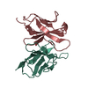
| ||||||||
|---|---|---|---|---|---|---|---|---|---|
| 1 |
| ||||||||
| Unit cell |
|
-Components
| #1: Antibody | Mass: 12223.854 Da / Num. of mol.: 1 / Fragment: FV / Mutation: CHAIN L, A100C, CHAIN H, R44C Source method: isolated from a genetically manipulated source Source: (gene. exp.)  |
|---|---|
| #2: Antibody | Mass: 13248.775 Da / Num. of mol.: 1 / Fragment: FV / Mutation: CHAIN L, A100C, CHAIN H, R44C Source method: isolated from a genetically manipulated source Source: (gene. exp.) |
| #3: Water | ChemComp-HOH /  Mass: 18.015 Da / Num. of mol.: 180 / Source method: isolated from a natural source / Formula: H2O Mass: 18.015 Da / Num. of mol.: 180 / Source method: isolated from a natural source / Formula: H2O |
| Has protein modification | Y |
-Experimental details
-Experiment
| Experiment | Method: X-RAY DIFFRACTION / Number of used crystals: 1 |
|---|
- Sample preparation
| Crystal | Density Matthews: 2.6 Å3/Da / Density % sol: 54 % | |||||||||||||||||||||||||
|---|---|---|---|---|---|---|---|---|---|---|---|---|---|---|---|---|---|---|---|---|---|---|---|---|---|---|
| Crystal grow | pH: 7 / Details: 1.6M HEPES PH-7.0 | |||||||||||||||||||||||||
| Crystal | *PLUS | |||||||||||||||||||||||||
| Crystal grow | *PLUS Method: vapor diffusion, hanging drop | |||||||||||||||||||||||||
| Components of the solutions | *PLUS
|
-Data collection
| Diffraction | Mean temperature: 293 K |
|---|---|
| Diffraction source | Source: ROTATING ANODE / Type: RIGAKU RUH2R / Wavelength: 1.5418 |
| Detector | Type: SIEMENS / Detector: AREA DETECTOR / Date: Dec 1, 1995 |
| Radiation | Monochromator: GRAPHITE(002) / Monochromatic (M) / Laue (L): M / Scattering type: x-ray |
| Radiation wavelength | Wavelength: 1.5418 Å / Relative weight: 1 |
| Reflection | Resolution: 2.12→57.8 Å / Num. obs: 14289 / % possible obs: 92 % / Observed criterion σ(I): 0 / Redundancy: 6 % / Rsym value: 0.108 / Net I/σ(I): 6.8 |
| Reflection shell | Resolution: 2.12→2.2 Å / Redundancy: 3.4 % / Mean I/σ(I) obs: 1.5 / Rsym value: 0.2644 / % possible all: 58 |
| Reflection | *PLUS Num. all: 15524 / Observed criterion σ(I): 2 / Rmerge(I) obs: 0.066 |
- Processing
| Software |
| ||||||||||||||||||||||||||||||||||||||||||||||||||||||||||||
|---|---|---|---|---|---|---|---|---|---|---|---|---|---|---|---|---|---|---|---|---|---|---|---|---|---|---|---|---|---|---|---|---|---|---|---|---|---|---|---|---|---|---|---|---|---|---|---|---|---|---|---|---|---|---|---|---|---|---|---|---|---|
| Refinement | Method to determine structure: MOLECULAR REPLACEMENT Starting model: PDB ENTRY 1IGG Resolution: 2→8 Å / σ(F): 2 /
| ||||||||||||||||||||||||||||||||||||||||||||||||||||||||||||
| Refinement step | Cycle: LAST / Resolution: 2→8 Å
| ||||||||||||||||||||||||||||||||||||||||||||||||||||||||||||
| Refine LS restraints |
| ||||||||||||||||||||||||||||||||||||||||||||||||||||||||||||
| Xplor file |
|
