Resolution: 1.6→30 Å / Cor.coef. Fo:Fc: 0.955 / Cor.coef. Fo:Fc free: 0.931 / SU B: 1.171 / SU ML: 0.043 / Cross valid method: THROUGHOUT / ESU R: 0.094 / ESU R Free: 0.105 / Stereochemistry target values: MAXIMUM LIKELIHOOD Details: HYDROGENS HAVE BEEN ADDED IN THE RIDING POSITIONS THE LAST REFINEMENT ROUND WAS DONE AGAINST ALL DATA, RFREE VALUES ARE THE LAST RECORDED
Rfactor
Num. reflection
% reflection
Selection details
Rfree
0.214
1575
10.1 %
RANDOM
Rwork
0.172
-
-
-
obs
0.174
15620
99.8 %
-
Solvent computation
Ion probe radii: 0.8 Å / Shrinkage radii: 0.8 Å / VDW probe radii: 1.4 Å / Solvent model: BABINET MODEL WITH MASK
 Movie
Movie Controller

 Yorodumi
 Open data
 Basic information
 Components
 Keywords
 Function and homology information
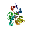
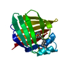
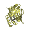
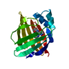
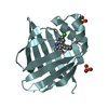
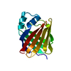
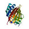
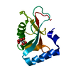
 ECHINOCOCCUS GRANULOSUS (invertebrata)
 X-RAY DIFFRACTION /
  Authors
 Citation
 Structure visualization
 Downloads & links
 Other downloads

 PDBj
PDBj


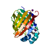
 Assembly
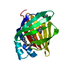


 Mass: 256.424 Da / Num. of mol.: 1 / Source method: obtained synthetically / Formula: C16H32O2 / Feature type: SUBJECT OF INVESTIGATION
 Mass: 18.015 Da / Num. of mol.: 143 / Source method: isolated from a natural source / Formula: H2O
 Sample preparation
 / Beamline: ID29 / Wavelength: 1.0038
  Processing