 Movie
Movie Controller


+ Open data

- Basic information
| Entry | Database: PDB / ID: 1a18 | |||||||||
|---|---|---|---|---|---|---|---|---|---|---|
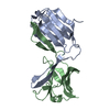
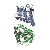
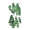
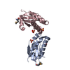
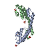
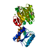
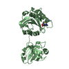
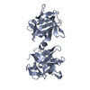
| Title | PHENANTHROLINE MODIFIED MURINE ADIPOCYTE LIPID BINDING PROTEIN | |||||||||
 Components Components | ADIPOCYTE LIPID BINDING PROTEIN | |||||||||
 Keywords Keywords | FATTY ACID BINDING PROTEIN / TRANSPORT / PHOSPHORYLATION | |||||||||
| Function / homology |  Function and homology information Function and homology informationlipase activator activity / Triglyceride catabolism / lipid droplet formation / triglyceride catabolic process / hormone receptor binding / long-chain fatty acid transmembrane transporter activity / long-chain fatty acid binding / cellular response to lithium ion / white fat cell differentiation / long-chain fatty acid transport ...lipase activator activity / Triglyceride catabolism / lipid droplet formation / triglyceride catabolic process / hormone receptor binding / long-chain fatty acid transmembrane transporter activity / long-chain fatty acid binding / cellular response to lithium ion / white fat cell differentiation / long-chain fatty acid transport / fatty acid transport / lipid droplet / brown fat cell differentiation / fatty acid binding / cholesterol homeostasis / response to bacterium / fatty acid metabolic process / cellular response to tumor necrosis factor / positive regulation of inflammatory response / positive regulation of cold-induced thermogenesis / negative regulation of DNA-templated transcription / positive regulation of cell population proliferation / nucleoplasm / nucleus / cytoplasm / cytosol Similarity search - Function | |||||||||
| Biological species |  | |||||||||
| Method |  X-RAY DIFFRACTION / MOLECULAR REPLACEMENT / Resolution: 2.4 Å X-RAY DIFFRACTION / MOLECULAR REPLACEMENT / Resolution: 2.4 Å | |||||||||
 Authors Authors | Ory, J. / Mazhary, A. / Kuang, H. / Davies, R. / Distefano, M. / Banaszak, L. | |||||||||
 Citation Citation | Journal: Protein Eng. / Year: 1998 Title: Structural characterization of two synthetic catalysts based on adipocyte lipid-binding protein. Authors: Ory, J.J. / Mazhary, A. / Kuang, H. / Davies, R.R. / Distefano, M.D. / Banaszak, L.J. | |||||||||
| History |
|
- Structure visualization
| Structure viewer | Molecule: MolmilJmol/JSmol |
|---|
- Downloads & links
-Download
| PDBx/mmCIF format | 1a18.cif.gz | 34.3 KB | Display | PDBx/mmCIF format |
|---|---|---|---|---|
| PDB format | pdb1a18.ent.gz | 26.2 KB | Display | PDB format |
| PDBx/mmJSON format | 1a18.json.gz | Tree view | PDBx/mmJSON format | |
| Others |  Other downloads Other downloads |
-Validation report
| Arichive directory | https://data.pdbj.org/pub/pdb/validation_reports/a1/1a18ftp://data.pdbj.org/pub/pdb/validation_reports/a1/1a18 | HTTPS FTP |
|---|
-Related structure data
| Related structure data |  1a2dC  1libS S: Starting model for refinement C: citing same article ( |
|---|---|
| Similar structure data |


-Links
 PDBj
PDBj

- Assembly
| Deposited unit | 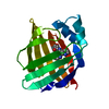
| ||||||||
|---|---|---|---|---|---|---|---|---|---|
| 1 | 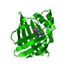
| ||||||||
| Unit cell |
| ||||||||
| Components on special symmetry positions |
|
-Components
| #1: Protein | Mass: 14774.931 Da / Num. of mol.: 1 Source method: isolated from a genetically manipulated source Details: PROTEIN MODIFIED BY REACTION WITH IODOACETAMIDO-1,10-PHENANTHROLINE Source: (gene. exp.) Description: SEE DAVIES AND DISTEFANO, JACS 119, 11643- 11652 (1977) Cell: ADIPOCYTE / Production host:  |
|---|---|
| #2: Water | ChemComp-HOH /  Mass: 18.015 Da / Num. of mol.: 38 / Source method: isolated from a natural source / Formula: H2O Mass: 18.015 Da / Num. of mol.: 38 / Source method: isolated from a natural source / Formula: H2O |
-Experimental details
-Experiment
| Experiment | Method: X-RAY DIFFRACTION / Number of used crystals: 1 |
|---|
- Sample preparation
| Crystal | Density Matthews: 2.2 Å3/Da / Density % sol: 45.6 % | ||||||||||||||||||||||||||||||
|---|---|---|---|---|---|---|---|---|---|---|---|---|---|---|---|---|---|---|---|---|---|---|---|---|---|---|---|---|---|---|---|
| Crystal grow | pH: 6.5 / Details: pH 6.5 | ||||||||||||||||||||||||||||||
| Crystal | *PLUS | ||||||||||||||||||||||||||||||
| Crystal grow | *PLUS pH: 7.5 / Method: vapor diffusion, hanging drop | ||||||||||||||||||||||||||||||
| Components of the solutions | *PLUS
|
-Data collection
| Diffraction | Mean temperature: 298 K |
|---|---|
| Diffraction source | Source: ROTATING ANODE / Type: RIGAKU RUH2R / Wavelength: 1.5418 |
| Detector | Type: SIEMENS / Detector: AREA DETECTOR / Date: Jun 1, 1996 |
| Radiation | Monochromator: GRAPHITE(002) / Monochromatic (M) / Laue (L): M / Scattering type: x-ray |
| Radiation wavelength | Wavelength: 1.5418 Å / Relative weight: 1 |
| Reflection | Resolution: 2.4→25 Å / Num. obs: 7640 / % possible obs: 91.7 % / Observed criterion σ(I): 0 / Redundancy: 4.5 % / Rsym value: 0.078 / Net I/σ(I): 13.1 |
| Reflection shell | Resolution: 2.37→2.52 Å / Redundancy: 2 % / Mean I/σ(I) obs: 2.3 / Rsym value: 0.325 / % possible all: 62.4 |
| Reflection | *PLUS Num. measured all: 34268 / Rmerge(I) obs: 0.079 |
| Reflection shell | *PLUS Rmerge(I) obs: 0.325 |
- Processing
| Software |
| ||||||||||||||||||||
|---|---|---|---|---|---|---|---|---|---|---|---|---|---|---|---|---|---|---|---|---|---|
| Refinement | Method to determine structure: MOLECULAR REPLACEMENT Starting model: PDB ENTRY 1LIB Resolution: 2.4→25 Å / Rfactor Rfree error: 0.0124 / Data cutoff high absF: 1000000 / Data cutoff low absF: 0.001 / Isotropic thermal model: RESTRAINED / Cross valid method: THROUGHOUT / σ(F): 1
| ||||||||||||||||||||
| Refinement step | Cycle: LAST / Resolution: 2.4→25 Å
| ||||||||||||||||||||
| LS refinement shell | Resolution: 2.4→2.51 Å / Rfactor Rfree error: 0.05 / Total num. of bins used: 8
| ||||||||||||||||||||
| Xplor file |
| ||||||||||||||||||||
| Software | *PLUS Name: X-PLOR / Version: 3.843C / Classification: refinement | ||||||||||||||||||||
| Refine LS restraints | *PLUS
|
