 Movie
Movie Controller

[English] 日本語
 Yorodumi
- PDB-6k6r: Crystal structure of Saccharomyces cerevisiae single domain sulfu... -
+ Open data

- Basic information
| Entry | Database: PDB / ID: 6k6r | ||||||
|---|---|---|---|---|---|---|---|
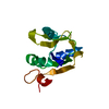
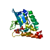
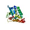
| Title | Crystal structure of Saccharomyces cerevisiae single domain sulfurtranferase RDL2 | ||||||
 Components Components | Thiosulfate sulfurtransferase RDL2, mitochondrial | ||||||
 Keywords Keywords | TRANSFERASE / thiosulfate:cyanide sulfurtransferase / Rhodanese / Saccharomyces cerevisiae | ||||||
| Function / homology |  Function and homology information Function and homology informationthiosulfate sulfurtransferase / sulfur compound metabolic process / thiosulfate-cyanide sulfurtransferase activity / mitochondrion Similarity search - Function | ||||||
| Biological species |  | ||||||
| Method |  X-RAY DIFFRACTION / SYNCHROTRON / MOLECULAR REPLACEMENT / Resolution: 2.471 Å X-RAY DIFFRACTION / SYNCHROTRON / MOLECULAR REPLACEMENT / Resolution: 2.471 Å | ||||||
 Authors Authors | Li, H.J. / Wang, Q.D. | ||||||
| Funding support |  China, 1items China, 1items
| ||||||
 Citation Citation | Journal: Antioxidants / Year: 2021 Title: Saccharomyces cerevisiaeRhodanese RDL2 Uses the ArgResidue of the Active-Site Loop for Thiosulfate Decomposition Authors: Wang, Q.D. / Li, H. / Xia, Y. / Xun, L. / Li, H.J. | ||||||
| History |
|
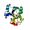
- Structure visualization
| Structure viewer | Molecule: MolmilJmol/JSmol |
|---|
- Downloads & links
-Download
| PDBx/mmCIF format | 6k6r.cif.gz | 63.6 KB | Display | PDBx/mmCIF format |
|---|---|---|---|---|
| PDB format | pdb6k6r.ent.gz | 45.6 KB | Display | PDB format |
| PDBx/mmJSON format | 6k6r.json.gz | Tree view | PDBx/mmJSON format | |
| Others |  Other downloads Other downloads |
-Validation report
| Arichive directory | https://data.pdbj.org/pub/pdb/validation_reports/k6/6k6rftp://data.pdbj.org/pub/pdb/validation_reports/k6/6k6r | HTTPS FTP |
|---|
-Related structure data
| Related structure data |  3d1pS S: Starting model for refinement |
|---|---|
| Similar structure data |


-Links
 PDBj
PDBj- Assembly
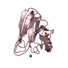
| Deposited unit | 
| ||||||||||||
|---|---|---|---|---|---|---|---|---|---|---|---|---|---|
| 1 | 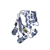
| ||||||||||||
| 2 | 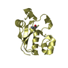
| ||||||||||||
| Unit cell |
| ||||||||||||
| Components on special symmetry positions |
|
-Components
| #1: Protein | Mass: 16758.461 Da / Num. of mol.: 2 Source method: isolated from a genetically manipulated source Source: (gene. exp.) Strain: ATCC 204508 / S288c / Gene: RDL2, AIM42, FMP31, YOR286W Production host:  References: UniProt: Q08742, thiosulfate sulfurtransferase #2: Water | ChemComp-HOH / |  Mass: 18.015 Da / Num. of mol.: 67 / Source method: isolated from a natural source / Formula: H2O Mass: 18.015 Da / Num. of mol.: 67 / Source method: isolated from a natural source / Formula: H2OHas protein modification | Y | |
|---|
-Experimental details
-Experiment
| Experiment | Method: X-RAY DIFFRACTION / Number of used crystals: 1 |
|---|
- Sample preparation
| Crystal | Density Matthews: 2.61 Å3/Da / Density % sol: 52.89 % |
|---|---|
| Crystal grow | Temperature: 289.15 K / Method: vapor diffusion, hanging drop / pH: 7 / Details: 3.5M Ammonium citrate |
-Data collection
| Diffraction | Mean temperature: 100 K / Serial crystal experiment: N |
|---|---|
| Diffraction source | Source: SYNCHROTRON / Site: SSRF / Beamline: BL17U / Wavelength: 0.9791 Å |
| Detector | Type: DECTRIS PILATUS3 6M / Detector: PIXEL / Date: Oct 13, 2018 |
| Radiation | Protocol: SINGLE WAVELENGTH / Monochromatic (M) / Laue (L): M / Scattering type: x-ray |
| Radiation wavelength | Wavelength: 0.9791 Å / Relative weight: 1 |
| Reflection | Resolution: 2.47→50 Å / Num. obs: 11448 / % possible obs: 100 % / Redundancy: 13.1 % / CC1/2: 0.99 / Net I/σ(I): 24.69 |
| Reflection shell | Resolution: 2.47→2.51 Å |
- Processing
| Software |
| ||||||||||||||||
|---|---|---|---|---|---|---|---|---|---|---|---|---|---|---|---|---|---|
| Refinement | Method to determine structure: MOLECULAR REPLACEMENT Starting model: 3D1P Resolution: 2.471→27.595 Å / Cross valid method: FREE R-VALUE
| ||||||||||||||||
| Refinement step | Cycle: LAST / Resolution: 2.471→27.595 Å
|
