Mass: 18.015 Da / Num. of mol.: 237 / Source method: isolated from a natural source / Formula: H2O
Nonpolymer details
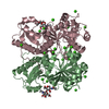
THE CRITERION TO ASSIGN A SOLVENT PEAK AS A POTASSIUM ION WAS THE ISOTROPIC TEMPERATURE FACTOR UPON ...THE CRITERION TO ASSIGN A SOLVENT PEAK AS A POTASSIUM ION WAS THE ISOTROPIC TEMPERATURE FACTOR UPON REFINEMENT IN X-PLOR. IF B REFINED TO 2.0, THE MINIMUM POSSIBLE IN X-PLOR, THEN THE SOLVENT PEAK WAS ASSIGNED AS POTASSIUM ION (PRESENT IN THE MOTHER LIQUOR) INSTEAD OF A WATER MOLECULE. FOR SIX SOLVENT PEAKS THE REFINEMENT PROCEEDED SMOOTHLY WHEN ASSIGNED TO BE POTASSIUM IONS.
-
Experimental details
-
Experiment
Experiment
Method: X-RAY DIFFRACTION / Number of used crystals: 1
-
Sample preparation
Crystal
Density Matthews: 2.24 Å3/Da / Density % sol: 55 %
Resolution: 1.9→30 Å / Num. obs: 9760 / % possible obs: 99 % / Observed criterion σ(I): 0
Reflection
*PLUS
Rmerge(I) obs: 0.04
-
Processing
Software
Name
Version
Classification
X-PLOR
3.1
modelbuilding
X-PLOR
3.1
refinement
XENGEN
datareduction
XENGEN
datascaling
X-PLOR
3.1
phasing
Refinement
Method to determine structure: SIR, NATIVE ANOMALOUS SCATTERING SOFTWARE USED : NULL STARTING MODEL FOR MOLECULAR REPLACEMENT: NULL Resolution: 1.9→30 Å / σ(F): 0
In the structure databanks used in Yorodumi, some data are registered as the other names, "COVID-19 virus" and "2019-nCoV". Here are the details of the virus and the list of structure data.
Jan 31, 2019. EMDB accession codes are about to change! (news from PDBe EMDB page)
EMDB accession codes are about to change! (news from PDBe EMDB page)
The allocation of 4 digits for EMDB accession codes will soon come to an end. Whilst these codes will remain in use, new EMDB accession codes will include an additional digit and will expand incrementally as the available range of codes is exhausted. The current 4-digit format prefixed with “EMD-” (i.e. EMD-XXXX) will advance to a 5-digit format (i.e. EMD-XXXXX), and so on. It is currently estimated that the 4-digit codes will be depleted around Spring 2019, at which point the 5-digit format will come into force.
The EM Navigator/Yorodumi systems omit the EMD- prefix.
Related info.:Q: What is EMD? / ID/Accession-code notation in Yorodumi/EM Navigator
Yorodumi is a browser for structure data from EMDB, PDB, SASBDB, etc.
This page is also the successor to EM Navigator detail page, and also detail information page/front-end page for Omokage search.
The word "yorodu" (or yorozu) is an old Japanese word meaning "ten thousand". "mi" (miru) is to see.
Related info.:EMDB / PDB / SASBDB / Comparison of 3 databanks / Yorodumi Search / Aug 31, 2016. New EM Navigator & Yorodumi / Yorodumi Papers / Jmol/JSmol / Function and homology information / Changes in new EM Navigator and Yorodumi
 Movie
Movie Controller


 Open data
 Basic information
 Components
 Keywords
 Function and homology information
 Haloarcula marismortui (Halophile)
 X-RAY DIFFRACTION /
  Authors
 Citation
 Structure visualization
 Downloads & links
 Other downloads
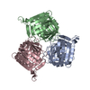


 PDBj
PDBj


 Assembly

 Mass: 39.098 Da / Num. of mol.: 6 / Source method: obtained synthetically / Formula: K
 Mass: 175.820 Da / Num. of mol.: 1 / Source method: obtained synthetically / Formula: Fe2S2
 Mass: 18.015 Da / Num. of mol.: 237 / Source method: isolated from a natural source / Formula: H2O
 Sample preparation
 Processing