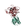
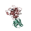
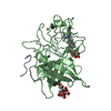
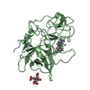
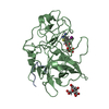
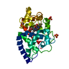
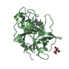
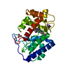
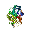
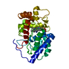
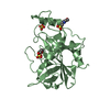
登録情報 データベース : PDB / ID : 1o5bタイトル Dissecting and Designing Inhibitor Selectivity Determinants at the S1 site Using an Artificial Ala190 Protease (Ala190 uPA) (Urokinase-type plasminogen activator) x 2 キーワード / / / / / / / / / 機能・相同性 分子機能 ドメイン・相同性 構成要素
/ / / / / / / / / / / / / / / / / / / / / / / / / / / / / / / / / / / / / / / / / / / / / / / / / / / / / / / / / / / / / / / / / / / / / / / / / / / / / / / / / / 生物種 Homo sapiens (ヒト)手法 / / 解像度 : 1.85 Å データ登録者 Katz, B.A. / Luong, C. / Ho, J.D. / Somoza, J.R. / Gjerstad, E. / Tang, J. / Williams, S.R. / Verner, E. / Mackman, R.L. / Young, W.B. ...Katz, B.A. / Luong, C. / Ho, J.D. / Somoza, J.R. / Gjerstad, E. / Tang, J. / Williams, S.R. / Verner, E. / Mackman, R.L. / Young, W.B. / Sprengeler, P.A. / Chan, H. / Mortara, K. / Janc, J.W. / McGrath, M.E. ジャーナル : J.Mol.Biol. / 年 : 2004タイトル : Dissecting and designing inhibitor selectivity determinants at the S1 site using an artificial Ala190 protease (Ala190 uPA)著者 : Katz, B.A. / Luong, C. / Ho, J.D. / Somoza, J.R. / Gjerstad, E. / Tang, J. / Williams, S.R. / Verner, E. / Mackman, R.L. / Young, W.B. / Sprengeler, P.A. / Chan, H. / Mortara, K. / Janc, J.W. / McGrath, M.E. 履歴 登録 2003年9月9日 登録サイト / 処理サイト 改定 1.0 2004年9月21日 Provider / タイプ 改定 1.1 2008年4月26日 Group 改定 1.2 2011年7月13日 Group 改定 1.3 2021年10月27日 Group / Derived calculations / カテゴリ / struct_ref_seq_dif / struct_siteItem _database_2.pdbx_DOI / _database_2.pdbx_database_accession ... _database_2.pdbx_DOI / _database_2.pdbx_database_accession / _struct_ref_seq_dif.details / _struct_site.pdbx_auth_asym_id / _struct_site.pdbx_auth_comp_id / _struct_site.pdbx_auth_seq_id 改定 1.4 2023年12月27日 Group / カテゴリ / chem_comp_bond改定 1.5 2024年11月20日 Group カテゴリ / pdbx_modification_feature
すべて表示 表示を減らす
 ムービー
ムービー コントローラー


 データを開く
 基本情報
基本情報 要素
要素 キーワード
キーワード 機能・相同性情報
 Homo sapiens (ヒト)
 X線回折 /
  データ登録者
 引用
引用 構造の表示
構造の表示 ダウンロードとリンク
 その他のダウンロード


 PDBj
PDBj


 集合体
集合体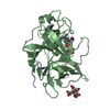
 Pichia pastoris (菌類) / 参照: UniProt: P00749, u-plasminogen activator
 分子量: 303.143 Da / 分子数: 1 / 由来タイプ: 合成 / 式: C9H8IN2S
 分子量: 192.124 Da / 分子数: 3 / 由来タイプ: 合成 / 式: C6H8O7
 分子量: 18.015 Da / 分子数: 319 / 由来タイプ: 天然 / 式: H2O
 試料調製
試料調製 解析
解析