 Movie
Movie Controller

[English] 日本語
 Yorodumi
- PDB-1o5e: Dissecting and Designing Inhibitor Selectivity Determinants at th... -
+ Open data

- Basic information
| Entry | Database: PDB / ID: 1o5e | ||||||
|---|---|---|---|---|---|---|---|
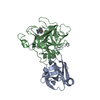
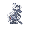
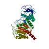
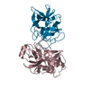
| Title | Dissecting and Designing Inhibitor Selectivity Determinants at the S1 site Using an Artificial Ala190 Protease (Ala190 uPA) | ||||||
 Components Components | (Serine protease hepsin) x 2 | ||||||
 Keywords Keywords | serine protease / hydrolase / srcr / scavenger receptor cysteine-rich domain / serine protease Ala190 uPA / S1 site / selectivity / conserved water displacement hydrogen bond deficit / trypsin / thrombin / hepsin / factor VIIa | ||||||
| Function / homology |  Function and homology information Function and homology informationhepsin / pilomotor reflex / positive regulation of thyroid hormone generation / Signaling by MST1 / serine-type exopeptidase activity / positive regulation of plasminogen activation / basement membrane disassembly / response to thyroid hormone / cochlea morphogenesis / MET Receptor Activation ...hepsin / pilomotor reflex / positive regulation of thyroid hormone generation / Signaling by MST1 / serine-type exopeptidase activity / positive regulation of plasminogen activation / basement membrane disassembly / response to thyroid hormone / cochlea morphogenesis / MET Receptor Activation / detection of mechanical stimulus involved in sensory perception of sound / positive regulation of hepatocyte proliferation / host-mediated activation of viral transcription / negative regulation of epithelial to mesenchymal transition / potassium ion transmembrane transport / serine-type peptidase activity / protein processing / negative regulation of epithelial cell proliferation / cell-cell junction / peptidase activity / regulation of cell shape / positive regulation of cell growth / apical plasma membrane / serine-type endopeptidase activity / neuronal cell body / positive regulation of gene expression / endoplasmic reticulum membrane / negative regulation of apoptotic process / cell surface / proteolysis / extracellular exosome / membrane / plasma membrane Similarity search - Function | ||||||
| Biological species |  Homo sapiens (human) Homo sapiens (human) | ||||||
| Method |  X-RAY DIFFRACTION / FOURIER SYNTHESIS / Resolution: 1.75 Å X-RAY DIFFRACTION / FOURIER SYNTHESIS / Resolution: 1.75 Å | ||||||
 Authors Authors | Katz, B.A. / Luong, C. / Ho, J.D. / Somoza, J.R. / Gjerstad, E. / Tang, J. / Williams, S.R. / Verner, E. / Mackman, R.L. / Young, W.B. ...Katz, B.A. / Luong, C. / Ho, J.D. / Somoza, J.R. / Gjerstad, E. / Tang, J. / Williams, S.R. / Verner, E. / Mackman, R.L. / Young, W.B. / Sprengeler, P.A. / Chan, H. / Mortara, K. / Janc, J.W. / McGrath, M.E. | ||||||
 Citation Citation | Journal: J.Mol.Biol. / Year: 2004 Title: Dissecting and designing inhibitor selectivity determinants at the S1 site using an artificial Ala190 protease (Ala190 uPA). Authors: Katz, B.A. / Luong, C. / Ho, J.D. / Somoza, J.R. / Gjerstad, E. / Tang, J. / Williams, S.R. / Verner, E. / Mackman, R.L. / Young, W.B. / Sprengeler, P.A. / Chan, H. / Mortara, K. / Janc, J.W. / McGrath, M.E. | ||||||
| History |
|
- Structure visualization
| Structure viewer | Molecule: MolmilJmol/JSmol |
|---|
- Downloads & links
-Download
| PDBx/mmCIF format | 1o5e.cif.gz | 158.7 KB | Display | PDBx/mmCIF format |
|---|---|---|---|---|
| PDB format | pdb1o5e.ent.gz | 126 KB | Display | PDB format |
| PDBx/mmJSON format | 1o5e.json.gz | Tree view | PDBx/mmJSON format | |
| Others |  Other downloads Other downloads |
-Validation report
| Arichive directory | https://data.pdbj.org/pub/pdb/validation_reports/o5/1o5eftp://data.pdbj.org/pub/pdb/validation_reports/o5/1o5e | HTTPS FTP |
|---|
-Related structure data
| Related structure data |  1o5aC  1o5bC  1o5cC  1o5dC 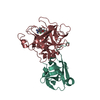 1o5fC  1o5gC C: citing same article ( |
|---|---|
| Similar structure data |


-Links
 PDBj
PDBj

- Assembly
| Deposited unit | 
| ||||||||||
|---|---|---|---|---|---|---|---|---|---|---|---|
| 1 |
| ||||||||||
| Unit cell |
|
-Components
| #1: Protein | Mass: 12522.299 Da / Num. of mol.: 1 / Fragment: light chain / Mutation: N67A Source method: isolated from a genetically manipulated source Source: (gene. exp.) Homo sapiens (human) / Gene: HPN OR TMPRSS1 / Production host:  Pichia pastoris (fungus) / Variant (production host): KM71 Pichia pastoris (fungus) / Variant (production host): KM71References: UniProt: P05981, Hydrolases; Acting on peptide bonds (peptidases); Serine endopeptidases |
|---|---|
| #2: Protein | Mass: 27573.111 Da / Num. of mol.: 1 / Fragment: heavy chain (catalytic domain) Source method: isolated from a genetically manipulated source Source: (gene. exp.) Homo sapiens (human) / Gene: HPN OR TMPRSS1 / Production host: Pichia pastoris (fungus) / Variant (production host): KM71References: UniProt: P05981, Hydrolases; Acting on peptide bonds (peptidases); Serine endopeptidases |
| #3: Chemical | ChemComp-132 /   Mass: 362.832 Da / Num. of mol.: 1 / Source method: obtained synthetically / Formula: C21H17ClN3O Mass: 362.832 Da / Num. of mol.: 1 / Source method: obtained synthetically / Formula: C21H17ClN3O |
| #4: Water | ChemComp-HOH /  Mass: 18.015 Da / Num. of mol.: 176 / Source method: isolated from a natural source / Formula: H2O Mass: 18.015 Da / Num. of mol.: 176 / Source method: isolated from a natural source / Formula: H2O |
| Has protein modification | Y |
-Experimental details
-Experiment
| Experiment | Method: X-RAY DIFFRACTION / Number of used crystals: 1 |
|---|
- Sample preparation
| Crystal | Density Matthews: 2.15 Å3/Da / Density % sol: 42.73 % |
|---|---|
| Crystal grow | Temperature: 290 K / Method: vapor diffusion / pH: 6.5 Details: 0.2 M ammonium fluoride, 20-25% PEG 3350, pH 7.6, vapor diffusion at 290 K Soak in synthetic mother liquor - CRA-10302 at pH 6.9., pH 6.5 |
-Data collection
| Diffraction | Mean temperature: 285 K |
|---|---|
| Diffraction source | Source: ROTATING ANODE / Type: RIGAKU RUH3R / Wavelength: 1.5418 |
| Detector | Type: RIGAKU RAXIS IV++ / Detector: IMAGE PLATE / Date: Nov 7, 2002 |
| Radiation | Protocol: SINGLE WAVELENGTH / Monochromatic (M) / Laue (L): M / Scattering type: x-ray |
| Radiation wavelength | Wavelength: 1.5418 Å / Relative weight: 1 |
| Reflection | Resolution: 1.75→25.8 Å / Num. all: 34620 / Num. obs: 29472 / % possible obs: 85.13 % / Observed criterion σ(I): 0 / Redundancy: 2.9 % / Rmerge(I) obs: 0.072 / Net I/σ(I): 5.3 |
| Reflection shell | Resolution: 1.75→1.83 Å / % possible obs: 30.7 % / Rmerge(I) obs: 0.435 / Num. unique all: 4235 |
- Processing
| Software |
| ||||||||||||||||||||
|---|---|---|---|---|---|---|---|---|---|---|---|---|---|---|---|---|---|---|---|---|---|
| Refinement | Method to determine structure: FOURIER SYNTHESIS / Resolution: 1.75→7 Å / Cross valid method: THROUGHOUT / σ(F): 0 / σ(I): 0 / Stereochemistry target values: X-PLOR force field Details: The first four residues of the light chain are not visible and are not included in the model. The loop comprising the sequence "RDPNSEE" between Phe_H97 and Asn_H99 is not visible and is not ...Details: The first four residues of the light chain are not visible and are not included in the model. The loop comprising the sequence "RDPNSEE" between Phe_H97 and Asn_H99 is not visible and is not included in the model. Residues simultaneously refined in two or more conformations are: Arg_L35, Val_L75, Arg_L79, Lys_L112, Leu_H41, Leu_H46, Gln_H73, Ser_H109, Pro_H185, Thr_H208, Lys_H224, Ser_H246, Gln_H254. Discretely disordered waters are HOH_212 and HOH_323. His_H40, HIS_H57, and His_H107 are doubly protonated. HIS_H91 is monoprotonated on the epsilon nitrogen No energy terms are included Og_Ser_H195, HOH_383, and O6' of the inhibitor. These atoms form a short hydrogen-bonding network.
| ||||||||||||||||||||
| Refinement step | Cycle: LAST / Resolution: 1.75→7 Å
| ||||||||||||||||||||
| Refine LS restraints |
|
