[English] 日本語
 Yorodumi
- PDB-1o1g: MOLECULAR MODELS OF AVERAGED RIGOR CROSSBRIDGES FROM TOMOGRAMS OF... -
+ Open data
- Basic information
| Entry | Database: PDB / ID: 1o1g | ||||||
|---|---|---|---|---|---|---|---|
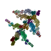
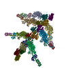
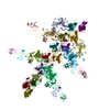
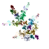
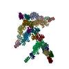
| Title | MOLECULAR MODELS OF AVERAGED RIGOR CROSSBRIDGES FROM TOMOGRAMS OF INSECT FLIGHT MUSCLE | ||||||
 Components Components |
| ||||||
 Keywords Keywords | CONTRACTILE PROTEIN / ACTIN-MYOSIN COMPLEX IN SITU IN MUSCLE | ||||||
| Function / homology |  Function and homology information Function and homology informationcontractile muscle fiber / Striated Muscle Contraction / myosin filament / myosin complex / myosin II complex / cytoskeletal motor activator activity / myosin heavy chain binding / microfilament motor activity / tropomyosin binding / actin filament bundle ...contractile muscle fiber / Striated Muscle Contraction / myosin filament / myosin complex / myosin II complex / cytoskeletal motor activator activity / myosin heavy chain binding / microfilament motor activity / tropomyosin binding / actin filament bundle / troponin I binding / filamentous actin / myofibril / mesenchyme migration / skeletal muscle myofibril / actin filament bundle assembly / striated muscle thin filament / skeletal muscle thin filament assembly / actin monomer binding / skeletal muscle tissue development / skeletal muscle fiber development / stress fiber / titin binding / actin filament polymerization / muscle contraction / actin filament / filopodium / Hydrolases; Acting on acid anhydrides; Acting on acid anhydrides to facilitate cellular and subcellular movement / calcium-dependent protein binding / actin filament binding / lamellipodium / cell body / calmodulin binding / hydrolase activity / protein domain specific binding / calcium ion binding / positive regulation of gene expression / magnesium ion binding / ATP binding / identical protein binding / cytoplasm Similarity search - Function | ||||||
| Biological species |   | ||||||
| Method | ELECTRON MICROSCOPY / electron tomography / negative staining / Resolution: 70 Å | ||||||
 Authors Authors | Chen, L.F. / Winkler, H. / Reedy, M.K. / Reedy, M.C. / Taylor, K.A. | ||||||
 Citation Citation |  Journal: J Struct Biol / Year: 2002 Journal: J Struct Biol / Year: 2002Title: Molecular modeling of averaged rigor crossbridges from tomograms of insect flight muscle. Authors: Li Fan Chen / Hanspeter Winkler / Michael K Reedy / Mary C Reedy / Kenneth A Taylor /  Abstract: Electron tomography, correspondence analysis, molecular model building, and real-space refinement provide detailed 3-D structures for in situ myosin crossbridges in the nucleotide-free state (rigor), ...Electron tomography, correspondence analysis, molecular model building, and real-space refinement provide detailed 3-D structures for in situ myosin crossbridges in the nucleotide-free state (rigor), thought to represent the end of the power stroke. Unaveraged tomograms from a 25-nm longitudinal section of insect flight muscle preserved native structural variation. Recurring crossbridge motifs that repeat every 38.7 nm along the actin filament were extracted from the tomogram and classified by correspondence analysis into 25 class averages, which improved the signal to noise ratio. Models based on the atomic structures of actin and of myosin subfragment 1 were rebuilt to fit 11 class averages. A real-space refinement procedure was applied to quantitatively fit the reconstructions and to minimize steric clashes between domains introduced during the fitting. These combined procedures show that no single myosin head structure can fit all the in situ crossbridges. The validity of the approach is supported by agreement of these atomic models with fluorescent probe data from vertebrate muscle as well as with data from regulatory light chain crosslinking between heads of smooth muscle heavy meromyosin when bound to actin. #1:  Journal: J.Struct.Biol. / Year: 2001 Journal: J.Struct.Biol. / Year: 2001Title: Real Space Refinement of Acto-Myosin Structures from Sectioned Muscle. Authors: Chen, L.F. / Blanc, E. / Chapman, M.S. / Taylor, K.A. #2:  Journal: Ultramicroscopy / Year: 1999 Journal: Ultramicroscopy / Year: 1999Title: Multivariate Statistical Analysis of Three-Dimensional Cross-Bridge Motifs in Insect Flight Muscle Authors: Winkler, H. / Taylor, K.A. #3:  Journal: J.Struct.Biol. / Year: 1997 Journal: J.Struct.Biol. / Year: 1997Title: The Use of Electron Tomography for Structural Analysis of Disordered Protein Arrays. Authors: Taylor, K.A. / Tang, J. / Cheng, Y. / Winkler, H. | ||||||
| History |
| ||||||
| Remark 999 | SEQUENCE Since the models used to fit the data are not from the same source, the DBREF and the ...SEQUENCE Since the models used to fit the data are not from the same source, the DBREF and the SEQADV remarks were suppressed. |
- Structure visualization
| Movie |
 Movie viewer Movie viewer |
|---|---|
| Structure viewer | Molecule:  Molmil Molmil Jmol/JSmol Jmol/JSmol |
- Downloads & links
- Download
| PDBx/mmCIF format |  1o1g.cif.gz 1o1g.cif.gz | 2 MB | Display |  PDBx/mmCIF format PDBx/mmCIF format |
|---|---|---|---|---|
| PDB format |  pdb1o1g.ent.gz pdb1o1g.ent.gz | 1.6 MB | Display |  PDB format PDB format |
| PDBx/mmJSON format |  1o1g.json.gz 1o1g.json.gz | Tree view |  PDBx/mmJSON format PDBx/mmJSON format | |
| Others |  Other downloads Other downloads |
-Validation report
| Arichive directory |  https://data.pdbj.org/pub/pdb/validation_reports/o1/1o1g https://data.pdbj.org/pub/pdb/validation_reports/o1/1o1g ftp://data.pdbj.org/pub/pdb/validation_reports/o1/1o1g ftp://data.pdbj.org/pub/pdb/validation_reports/o1/1o1g | HTTPS FTP |
|---|
-Related structure data
| Related structure data |  1001MK  1m8qC  1mvwC  1o18C  1o19C  1o1aC  1o1bC  1o1cC  1o1dC  1o1eC  1o1fC |
|---|---|
| Similar structure data |
- Links
Links
- Assembly
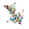
| Deposited unit | 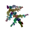
|
|---|---|
| 1 |
|
- Components
| #1: Protein | Mass: 96625.484 Da / Num. of mol.: 6 / Source method: isolated from a natural source Details: ORGANISM FROM WHICH THE MYOSIN FOR THE CRYSTAL STRUCTURE THAT PROVIDED 2MYS WAS OBTAINED Source: (natural)  #2: Protein | Mass: 16063.036 Da / Num. of mol.: 6 / Source method: isolated from a natural source Details: ORGANISM FROM WHICH THE MYOSIN FOR THE CRYSTAL STRUCTURE THAT PROVIDED 2MYS WAS OBTAINED Source: (natural)  #3: Protein | Mass: 16063.926 Da / Num. of mol.: 6 / Source method: isolated from a natural source Details: ORGANISM FROM WHICH THE MYOSIN FOR THE CRYSTAL STRUCTURE THAT PROVIDED 2MYS WAS OBTAINED Source: (natural)  #4: Protein | Mass: 41862.613 Da / Num. of mol.: 14 / Source method: isolated from a natural source Details: ORGANISM FROM WHICH THE ACTIN THAT PROVIDED THE CRYSTAL STRUCTURE FOR 1ATN WAS OBTAINED Source: (natural)  |
|---|
-Experimental details
-Experiment
| Experiment | Method: ELECTRON MICROSCOPY |
|---|---|
| EM experiment | Aggregation state: TISSUE / 3D reconstruction method: electron tomography |
- Sample preparation
| Component | Name: INSECT FLIGHT MUSCLE OF THE LARGE WATER BUG LETHOCERUS MAXIMUS Type: TISSUE Details: DORSAL LONGITUDINAL FLIGHT MUSCLES OF THE LARGE WATERBUG, LETHOCERUS MAXIMUS WERE GLYCERINATED IN THE DISSECTED THORAX AND STORED AT -20 DEGREES C. FIXATION INVOLVED SEQUENTIAL TANNIC ACID- ...Details: DORSAL LONGITUDINAL FLIGHT MUSCLES OF THE LARGE WATERBUG, LETHOCERUS MAXIMUS WERE GLYCERINATED IN THE DISSECTED THORAX AND STORED AT -20 DEGREES C. FIXATION INVOLVED SEQUENTIAL TANNIC ACID-GLUTARALDEHYDE (0.2% AND 2%), COLD 1% OSO4 AT PH 6.0, 1% URANYL ACETATE BLOCK STAINING AND ARALDITE 506 EMGEDDING. THIN SECTIONS WERE STAINED WITH A SEQUENCE OF PERMANGANATE AND LEAD. SECTION WERE ~25 NM THICK. MODELS ARE BUILT TO FIT THE THIN, ACTIN CONTAINING FILAMENTS LABELED WITH ENDOGENOUS MYOSIN IN THE RIGOR STATE. |
|---|---|
| Specimen | Embedding applied: YES / Shadowing applied: NO / Staining applied: YES / Vitrification applied: NO |
| EM staining | Type: NEGATIVE / Material: Osmium tetroxide, Uranyl Acetate |
| Vitrification | Details: NO VITRIFICATION. SAMPLES WERE VIEWED AT ROOM TEMPERATURE. |
- Electron microscopy imaging
| Microscopy | Model: FEI/PHILIPS EM400 |
|---|---|
| Electron gun | Electron source: TUNGSTEN HAIRPIN / Accelerating voltage: 100 kV / Illumination mode: FLOOD BEAM |
| Electron lens | Mode: BRIGHT FIELD / Nominal magnification: 17000 X |
| Image recording | Film or detector model: KODAK SO-163 FILM |
| Image scans | Num. digital images: 36 |
| Radiation | Protocol: SINGLE WAVELENGTH / Monochromatic (M) / Laue (L): M |
| Radiation wavelength | Relative weight: 1 |
- Processing
| EM software |
| |||||||||||||||||||||
|---|---|---|---|---|---|---|---|---|---|---|---|---|---|---|---|---|---|---|---|---|---|---|
| Symmetry | Point symmetry: C1 (asymmetric) | |||||||||||||||||||||
| 3D reconstruction | Method: DUAL AXIS TILT SERIES ELECTRON TOMOGRAPHY / Resolution: 70 Å / Nominal pixel size: 15.5 Å Magnification calibration: INDICATED INSTRUMENT MAGNIFICATION Details: 3-D MOTIFS WERE IDENTIFIED IN THE TOMOGRAM BY FIRST PRODUCING A CROSS CORRELATION MAP FROM WHICH PEAK COORDINATES WERE DETERMINED FROM THEIR CENTER OF GRAVITY. WE DEFINE A 3-D MOTIF AS ONE, ...Details: 3-D MOTIFS WERE IDENTIFIED IN THE TOMOGRAM BY FIRST PRODUCING A CROSS CORRELATION MAP FROM WHICH PEAK COORDINATES WERE DETERMINED FROM THEIR CENTER OF GRAVITY. WE DEFINE A 3-D MOTIF AS ONE, ENTIRE 38.7 NM CROSSBRIDGE REPEAT ALONG ACTIN. THESE MOTIFS USUALLY CONTAIN AT LEAST FOUR MYOSIN HEADS IN TWO PAIRED CROSSBRIDGES (SINGLE CHEVRONS) AND SOMETIMES CONTAIN AS MANY AS SIX MYOSIN HEADS IN FOUR PAIRED CROSSBRIDGES (DOUBLE CHEVRONS). THE REFERENCE FOR THE ANALYSIS WAS SELECTED TO BE CENTERED BETWEEN SUCCESSIVE TROPONIN DENSITIES WHICH COULD BE IDENTIFIED FROM THE IN-PLANE PROJECTION. THE INDIVIDUAL CROSSBRIDGE MOTIFS WERE THEN SUBJECTED TO MULTIVARIATE STATISTICAL ANALYSIS TO IDENTIFY CLUSTERS OF MOTIFS SHOWING SIMILAR CROSSBRIDGE STRUCTURE. THESE CLUSTERS FORMED THE CLASS AVERAGES. THE CHOICE OF STRUCTURE TO BE CLASSIFIED WAS DECIDED BY THE RESOLUTION AND THE LATER PROCESS OF MODEL BUILDING. AVERAGING WAS DONE ACCORDING TO THE HEIRARCHICAL ASCENDENT METHOD. THE RESOLUTION IN EACH OF THE CLASS AVERAGES WAS 7 NM BY THE SPECTRAL SIGNAL TO NOISE RATIO. Symmetry type: POINT | |||||||||||||||||||||
| Atomic model building | Protocol: RIGID BODY FIT / Space: REAL Target criteria: BEST CORRELATION COEFFICIENT AND FEWEST POOR CONTACTS Details: METHOD--INITIAL MODELS WERE FIT BY HAND USING O. THE FIT WAS THEN REFINED USING REAL SPACE REFINEMENT. REFINEMENT PROTOCOL--RIGID BODY | |||||||||||||||||||||
| Atomic model building |
| |||||||||||||||||||||
| Refinement | Highest resolution: 70 Å | |||||||||||||||||||||
| Refinement step | Cycle: LAST / Highest resolution: 70 Å
|
 Movie
Movie Controller


 PDBj
PDBj


