 Movie
Movie Controller

[English] 日本語
 Yorodumi
- PDB-1nof: THE FIRST CRYSTALLOGRAPHIC STRUCTURE OF A XYLANASE FROM GLYCOSYL ... -
+ Open data

- Basic information
| Entry | Database: PDB / ID: 1nof | ||||||
|---|---|---|---|---|---|---|---|
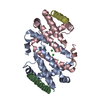
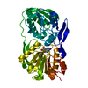
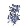
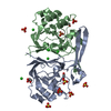
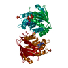
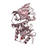
| Title | THE FIRST CRYSTALLOGRAPHIC STRUCTURE OF A XYLANASE FROM GLYCOSYL HYDROLASE FAMILY 5: IMPLICATIONS FOR CATALYSIS | ||||||
 Components Components | xylanase | ||||||
 Keywords Keywords | HYDROLASE / XYLANASE / GLYCOHYDROLASE FAMILY 5 / CARBOHYDRATE-BINDING MODULE / CATALYTIC DOMAIN | ||||||
| Function / homology |  Function and homology information Function and homology informationglucosylceramidase activity / sphingolipid metabolic process / xylan catabolic process / membrane Similarity search - Function | ||||||
| Biological species |  Erwinia chrysanthemi (bacteria) Erwinia chrysanthemi (bacteria) | ||||||
| Method |  X-RAY DIFFRACTION / MIR / Resolution: 1.42 Å X-RAY DIFFRACTION / MIR / Resolution: 1.42 Å | ||||||
 Authors Authors | Larson, S.B. / Day, J. / McPherson, A. / Barba De La Rosa, A.P. / Keen, N.T. | ||||||
 Citation Citation | Journal: Biochemistry / Year: 2003 Title: First crystallographic structure of a xylanase from glycoside hydrolase family 5: implications for catalysis. Authors: Larson, S.B. / Day, J. / Barba de la Rosa, A.P. / Keen, N.T. / McPherson, A. #1: Journal: Acta Crystallogr.,Sect.D / Year: 1997Title: Crystallization of Xylanase from Erwinia chrysanthemi: Influence of Heat and Polymeric Substrate Authors: Barba De La Rosa, A.P. / Day, J. / Larson, S.B. / Keen, N.T. / McPherson, A. #2: Journal: Mol.Plant Microbe Interact. / Year: 1996Title: Cloning and Characterization of a Xylanase Gene from Corn Strains of Erwinia chrysanthemi Authors: Keen, N.T. / Boyd, C. / Henrissat, B. | ||||||
| History |
|
- Structure visualization
| Structure viewer | Molecule: MolmilJmol/JSmol |
|---|
- Downloads & links
-Download
| PDBx/mmCIF format | 1nof.cif.gz | 178.1 KB | Display | PDBx/mmCIF format |
|---|---|---|---|---|
| PDB format | pdb1nof.ent.gz | 139.6 KB | Display | PDB format |
| PDBx/mmJSON format | 1nof.json.gz | Tree view | PDBx/mmJSON format | |
| Others |  Other downloads Other downloads |
-Validation report
| Arichive directory | https://data.pdbj.org/pub/pdb/validation_reports/no/1nofftp://data.pdbj.org/pub/pdb/validation_reports/no/1nof | HTTPS FTP |
|---|
-Related structure data
| Similar structure data |
|---|


-Links
 PDBj
PDBj- Assembly
| Deposited unit | 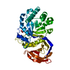
| ||||||||
|---|---|---|---|---|---|---|---|---|---|
| 1 |
| ||||||||
| Unit cell |
|
-Components
| #1: Protein | Mass: 42072.016 Da / Num. of mol.: 1 Source method: isolated from a genetically manipulated source Source: (gene. exp.) Erwinia chrysanthemi (bacteria) / Genus: Dickeya / Gene: xynA / Plasmid: pNTK136 / Production host: |
|---|---|
| #2: Chemical | ChemComp-ACT /   Mass: 59.044 Da / Num. of mol.: 1 / Source method: obtained synthetically / Formula: C2H3O2 Mass: 59.044 Da / Num. of mol.: 1 / Source method: obtained synthetically / Formula: C2H3O2 |
| #3: Water | ChemComp-HOH /  Mass: 18.015 Da / Num. of mol.: 522 / Source method: isolated from a natural source / Formula: H2O Mass: 18.015 Da / Num. of mol.: 522 / Source method: isolated from a natural source / Formula: H2O |
| Nonpolymer details | THE WATER MOLECULES HAVE BEEN NUMBERED TO REFLECT THEIR STATE 1001-1292 FIRST HYDRATION SHELL, FULL ...THE WATER MOLECULES HAVE BEEN NUMBERED TO REFLECT THEIR STATE 1001-1292 FIRST HYDRATION SHELL, FULL OCCUPANCY 2001-2048 FIRST HYDRATION SHELL, HALF OCCUPANCY 3001-4503 FIRST HYDRATION SHELL, DISORDERED |
| Sequence details | THE ENZYME USED IN THIS CRYSTALLOGRAPHIC STUDY IS A CLONE PRODUCT EXPRESSED IN E. COLI. UPON ...THE ENZYME USED IN THIS CRYSTALLOG |
-Experimental details
-Experiment
| Experiment | Method: X-RAY DIFFRACTION / Number of used crystals: 2 |
|---|
- Sample preparation
| Crystal | Density Matthews: 1.74 Å3/Da / Density % sol: 29.3 % |
|---|---|
| Crystal grow | Temperature: 291 K / Method: vapor diffusion / pH: 6.5 Details: 30% PEG 4000, 0.2 M AMMONIUM ACETATE, 0.1 M SODIUM CITRATE, pH 6.5, VAPOR DIFFUSION, temperature 291K |
| Crystal grow | *PLUS Details: Keen, N.T., (1996) Mol. Plant-Microbe Interact., 9, 651. |
-Data collection
| Diffraction | Mean temperature: 290 K |
|---|---|
| Diffraction source | Source: ROTATING ANODE / Type: RIGAKU RU200 / Wavelength: 1.5418 / Wavelength: 1.5418 Å |
| Detector | Type: SDMS / Detector: AREA DETECTOR / Date: Mar 1, 1996 |
| Radiation | Monochromator: SUPPER GRAPHITE / Protocol: SINGLE WAVELENGTH / Monochromatic (M) / Laue (L): M / Scattering type: x-ray |
| Radiation wavelength | Wavelength: 1.5418 Å / Relative weight: 1 |
| Reflection | Resolution: 1.42→44.57 Å / Num. all: 58915 / Num. obs: 58915 / % possible obs: 90.6 % / Observed criterion σ(F): 0 / Observed criterion σ(I): 0 / Redundancy: 3.51 % / Biso Wilson estimate: 12.67 Å2 / Rmerge(I) obs: 0.066 / Net I/σ(I): 10.17 |
| Reflection shell | Resolution: 1.42→1.52 Å / Redundancy: 1.91 % / Rmerge(I) obs: 0.132 / Mean I/σ(I) obs: 4.02 / Num. unique all: 8061 / % possible all: 55.8 |
| Reflection | *PLUS Num. measured all: 206521 / Rmerge(I) obs: 0.0656 |
| Reflection shell | *PLUS % possible obs: 61.9 % / Mean I/σ(I) obs: 3.14 |
- Processing
| Software |
| |||||||||||||||||||||||||||||||||
|---|---|---|---|---|---|---|---|---|---|---|---|---|---|---|---|---|---|---|---|---|---|---|---|---|---|---|---|---|---|---|---|---|---|---|
| Refinement | Method to determine structure: MIR / Resolution: 1.42→50 Å / Num. parameters: 30307 / Num. restraintsaints: 37937 / Cross valid method: FREE R THROUGHOUT / σ(F): 0 / Stereochemistry target values: ENGH AND HUBER
| |||||||||||||||||||||||||||||||||
| Solvent computation | Solvent model: MOEWS & KRETSINGER, J.MOL.BIOL.91(1973)201-228 | |||||||||||||||||||||||||||||||||
| Refine analyze | Luzzati coordinate error obs: 0.07 Å / Luzzati d res low obs: 2 Å / Num. disordered residues: 28 / Occupancy sum hydrogen: 2893.99 / Occupancy sum non hydrogen: 3389.15 | |||||||||||||||||||||||||||||||||
| Refinement step | Cycle: LAST / Resolution: 1.42→50 Å
| |||||||||||||||||||||||||||||||||
| Refine LS restraints |
| |||||||||||||||||||||||||||||||||
| Software | *PLUS Name: SHELXL / Version: 97 / Classification: refinement | |||||||||||||||||||||||||||||||||
| Refinement | *PLUS Lowest resolution: 9999 Å | |||||||||||||||||||||||||||||||||
| Solvent computation | *PLUS | |||||||||||||||||||||||||||||||||
| Displacement parameters | *PLUS | |||||||||||||||||||||||||||||||||
| Refine LS restraints | *PLUS
|
