 Movie
Movie Controller

[English] 日本語
 Yorodumi
- PDB-3v0g: Crystal structure of Ciona intestinalis voltage sensor-containing... -
+ Open data

- Basic information
| Entry | Database: PDB / ID: 3v0g | ||||||
|---|---|---|---|---|---|---|---|
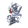
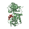
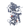
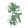
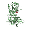
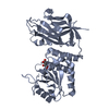
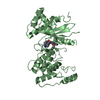
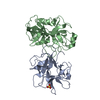
| Title | Crystal structure of Ciona intestinalis voltage sensor-containing phosphatase (Ci-VSP), residues 241-576(C363S), form III | ||||||
 Components Components | Voltage-sensor containing phosphatase | ||||||
 Keywords Keywords | HYDROLASE / PTP / C2 / Phosphatase | ||||||
| Function / homology |  Function and homology information Function and homology information | ||||||
| Biological species |  | ||||||
| Method |  X-RAY DIFFRACTION / SYNCHROTRON / MOLECULAR REPLACEMENT / Resolution: 1.6 Å X-RAY DIFFRACTION / SYNCHROTRON / MOLECULAR REPLACEMENT / Resolution: 1.6 Å | ||||||
 Authors Authors | Liu, L. / Kohout, S.C. / Xu, Q. / Muller, S. / Kimberlin, C. / Isacoff, E.Y. / Minor, D.L. | ||||||
 Citation Citation | Journal: Nat.Struct.Mol.Biol. / Year: 2012 Title: A glutamate switch controls voltage-sensitive phosphatase function. Authors: Liu, L. / Kohout, S.C. / Xu, Q. / Muller, S. / Kimberlin, C.R. / Isacoff, E.Y. / Minor, D.L. | ||||||
| History |
|
- Structure visualization
| Structure viewer | Molecule: MolmilJmol/JSmol |
|---|
- Downloads & links
-Download
| PDBx/mmCIF format | 3v0g.cif.gz | 296 KB | Display | PDBx/mmCIF format |
|---|---|---|---|---|
| PDB format | pdb3v0g.ent.gz | 237.4 KB | Display | PDB format |
| PDBx/mmJSON format | 3v0g.json.gz | Tree view | PDBx/mmJSON format | |
| Others |  Other downloads Other downloads |
-Validation report
| Arichive directory | https://data.pdbj.org/pub/pdb/validation_reports/v0/3v0gftp://data.pdbj.org/pub/pdb/validation_reports/v0/3v0g | HTTPS FTP |
|---|
-Related structure data
| Related structure data |  3v0dSC  3v0eC  3v0fC  3v0hC  3v0iC  3v0jC S: Starting model for refinement C: citing same article ( |
|---|---|
| Similar structure data |


-Links
 PDBj
PDBj


- Assembly
| Deposited unit | 
| ||||||||
|---|---|---|---|---|---|---|---|---|---|
| 1 | 
| ||||||||
| 2 | 
| ||||||||
| 3 | 
| ||||||||
| 4 | 
| ||||||||
| Unit cell |
|
-Components
| #1: Protein | Mass: 38827.984 Da / Num. of mol.: 4 / Fragment: unp residues 241-576 / Mutation: C363S Source method: isolated from a genetically manipulated source Source: (gene. exp.)  #2: Chemical | ChemComp-PO4 /   Mass: 94.971 Da / Num. of mol.: 9 / Source method: obtained synthetically / Formula: PO4 Mass: 94.971 Da / Num. of mol.: 9 / Source method: obtained synthetically / Formula: PO4#3: Water | ChemComp-HOH / |  Mass: 18.015 Da / Num. of mol.: 1169 / Source method: isolated from a natural source / Formula: H2O Mass: 18.015 Da / Num. of mol.: 1169 / Source method: isolated from a natural source / Formula: H2O |
|---|
-Experimental details
-Experiment
| Experiment | Method: X-RAY DIFFRACTION / Number of used crystals: 1 |
|---|
- Sample preparation
| Crystal | Density Matthews: 2.29 Å3/Da / Density % sol: 46.33 % |
|---|---|
| Crystal grow | Temperature: 277 K / Method: vapor diffusion, hanging drop / pH: 8 Details: 17.5-22.5% PEG 2000, 0.1 M ammonium dihydrophosphate, 0.1 M Tris-HCl,pH 8.0; crystals were dehydrated in 30% PEG 2000, 0.1 M ammonium dihydrophosphate, 0.1 M Tris-HCl,pH 8.0, VAPOR ...Details: 17.5-22.5% PEG 2000, 0.1 M ammonium dihydrophosphate, 0.1 M Tris-HCl,pH 8.0; crystals were dehydrated in 30% PEG 2000, 0.1 M ammonium dihydrophosphate, 0.1 M Tris-HCl,pH 8.0, VAPOR DIFFUSION, HANGING DROP, temperature 277K |
-Data collection
| Diffraction | Mean temperature: 100 K |
|---|---|
| Diffraction source | Source: SYNCHROTRON / Site: ALS  / Beamline: 8.3.1 / Wavelength: 1.1158 Å / Beamline: 8.3.1 / Wavelength: 1.1158 Å |
| Detector | Type: ADSC QUANTUM 315r / Detector: CCD / Date: Jan 28, 2010 / Details: Double Crystal Si(111) |
| Radiation | Monochromator: KHOZU Double flat crystal / Protocol: SINGLE WAVELENGTH / Monochromatic (M) / Laue (L): M / Scattering type: x-ray |
| Radiation wavelength | Wavelength: 1.1158 Å / Relative weight: 1 |
| Reflection | Resolution: 1.5→83.8 Å / Num. all: 220438 / Num. obs: 176659 / % possible obs: 80.5 % / Observed criterion σ(F): 2 / Observed criterion σ(I): 2 / Redundancy: 2.6 % / Rmerge(I) obs: 0.055 / Net I/σ(I): 15.3 |
| Reflection shell | Resolution: 1.5→1.5 Å / Redundancy: 1.4 % / Rmerge(I) obs: 0.29 / Mean I/σ(I) obs: 2.4 / % possible all: 83.1 |
- Processing
| Software |
| |||||||||||||||||||||||||||||||||||||||||||||||||||||||||||||||||
|---|---|---|---|---|---|---|---|---|---|---|---|---|---|---|---|---|---|---|---|---|---|---|---|---|---|---|---|---|---|---|---|---|---|---|---|---|---|---|---|---|---|---|---|---|---|---|---|---|---|---|---|---|---|---|---|---|---|---|---|---|---|---|---|---|---|---|
| Refinement | Method to determine structure: MOLECULAR REPLACEMENT Starting model: PDB: 3V0D Resolution: 1.6→83.71 Å / Cor.coef. Fo:Fc: 0.91 / Cor.coef. Fo:Fc free: 0.882 / SU B: 3.119 / SU ML: 0.111 / Cross valid method: THROUGHOUT / σ(F): 0 / ESU R: 0.162 / ESU R Free: 0.153 / Stereochemistry target values: MAXIMUM LIKELIHOOD
| |||||||||||||||||||||||||||||||||||||||||||||||||||||||||||||||||
| Solvent computation | Ion probe radii: 0.8 Å / Shrinkage radii: 0.8 Å / VDW probe radii: 1.4 Å / Solvent model: BABINET MODEL WITH MASK | |||||||||||||||||||||||||||||||||||||||||||||||||||||||||||||||||
| Displacement parameters | Biso mean: 24.766 Å2
| |||||||||||||||||||||||||||||||||||||||||||||||||||||||||||||||||
| Refinement step | Cycle: LAST / Resolution: 1.6→83.71 Å
| |||||||||||||||||||||||||||||||||||||||||||||||||||||||||||||||||
| Refine LS restraints |
| |||||||||||||||||||||||||||||||||||||||||||||||||||||||||||||||||
| LS refinement shell | Resolution: 1.6→1.642 Å / Total num. of bins used: 20
|
