 Movie
Movie Controller

[English] 日本語
 Yorodumi
- PDB-1nnd: Arginine 116 is Essential for Nucleic Acid Recognition by the Fin... -
+ Open data

- Basic information
| Entry | Database: PDB / ID: 1nnd | ||||||
|---|---|---|---|---|---|---|---|
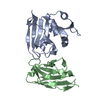
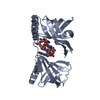
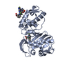
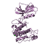
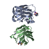
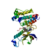
| Title | Arginine 116 is Essential for Nucleic Acid Recognition by the Fingers Domain of Moloney Murine Leukemia Virus Reverse Transcriptase | ||||||
 Components Components | Reverse Transcriptase | ||||||
 Keywords Keywords | TRANSFERASE / Nucleic Acid Binding / MMLV Reverse Transcriptase | ||||||
| Function / homology |  Function and homology information Function and homology informationretroviral 3' processing activity / host cell late endosome membrane / DNA catabolic process / Hydrolases; Acting on peptide bonds (peptidases); Aspartic endopeptidases / ribonuclease H / virion assembly / protein-DNA complex / viral genome integration into host DNA / establishment of integrated proviral latency / RNA-directed DNA polymerase ...retroviral 3' processing activity / host cell late endosome membrane / DNA catabolic process / Hydrolases; Acting on peptide bonds (peptidases); Aspartic endopeptidases / ribonuclease H / virion assembly / protein-DNA complex / viral genome integration into host DNA / establishment of integrated proviral latency / RNA-directed DNA polymerase / host multivesicular body / RNA-directed DNA polymerase activity / RNA-DNA hybrid ribonuclease activity / Transferases; Transferring phosphorus-containing groups; Nucleotidyltransferases / viral nucleocapsid / DNA recombination / DNA-directed DNA polymerase / structural constituent of virion / aspartic-type endopeptidase activity / Hydrolases; Acting on ester bonds / DNA-directed DNA polymerase activity / symbiont-mediated suppression of host gene expression / symbiont entry into host cell / host cell plasma membrane / proteolysis / DNA binding / RNA binding / zinc ion binding Similarity search - Function | ||||||
| Biological species |  Moloney murine leukemia virus Moloney murine leukemia virus | ||||||
| Method |  X-RAY DIFFRACTION / MOLECULAR REPLACEMENT / Resolution: 2.3 Å X-RAY DIFFRACTION / MOLECULAR REPLACEMENT / Resolution: 2.3 Å | ||||||
 Authors Authors | Crowther, R.L. / Remeta, D.P. / Minetti, C.A. / Das, D. / Montano, S.P. / Georgiadis, M.M. | ||||||
 Citation Citation | Journal: Proteins / Year: 2004 Title: Structural and energetic characterization of nucleic acid-binding to the fingers domain of Moloney murine leukemia virus reverse transcriptase Authors: Crowther, R.L. / Remeta, D.P. / Minetti, C.A. / Das, D. / Montano, S.P. / Georgiadis, M.M. | ||||||
| History |
|
- Structure visualization
| Structure viewer | Molecule: MolmilJmol/JSmol |
|---|
- Downloads & links
-Download
| PDBx/mmCIF format | 1nnd.cif.gz | 66 KB | Display | PDBx/mmCIF format |
|---|---|---|---|---|
| PDB format | pdb1nnd.ent.gz | 47.7 KB | Display | PDB format |
| PDBx/mmJSON format | 1nnd.json.gz | Tree view | PDBx/mmJSON format | |
| Others |  Other downloads Other downloads |
-Validation report
| Arichive directory | https://data.pdbj.org/pub/pdb/validation_reports/nn/1nndftp://data.pdbj.org/pub/pdb/validation_reports/nn/1nnd | HTTPS FTP |
|---|
-Related structure data
| Related structure data |  1qaiS S: Starting model for refinement |
|---|---|
| Similar structure data |


-Links
 PDBj
PDBj
- Assembly
| Deposited unit | 
| ||||||||||
|---|---|---|---|---|---|---|---|---|---|---|---|
| 1 |
| ||||||||||
| Unit cell |
|
-Components
| #1: Protein | Mass: 28848.170 Da / Num. of mol.: 1 / Fragment: MMLV Reverse Transcriptase / Mutation: R116A Source method: isolated from a genetically manipulated source Details: part of Pol polyprotein / Source: (gene. exp.) Moloney murine leukemia virus / Genus: Gammaretrovirus / Species: Murine leukemia virus / Gene: Reverse Transcriptase / Plasmid: pET15b / Production host:  |
|---|---|
| #2: Water | ChemComp-HOH /  Mass: 18.015 Da / Num. of mol.: 135 / Source method: isolated from a natural source / Formula: H2O Mass: 18.015 Da / Num. of mol.: 135 / Source method: isolated from a natural source / Formula: H2O |
-Experimental details
-Experiment
| Experiment | Method: X-RAY DIFFRACTION / Number of used crystals: 1 |
|---|
- Sample preparation
| Crystal | Density Matthews: 2.33 Å3/Da / Density % sol: 46.8 % | |||||||||||||||||||||||||||||||||||||||||||||||||
|---|---|---|---|---|---|---|---|---|---|---|---|---|---|---|---|---|---|---|---|---|---|---|---|---|---|---|---|---|---|---|---|---|---|---|---|---|---|---|---|---|---|---|---|---|---|---|---|---|---|---|
| Crystal grow | Temperature: 293 K / Method: vapor diffusion, hanging drop / pH: 7 Details: PEG 400, Magnesium Chloride, HEPES, pH 7.0, VAPOR DIFFUSION, HANGING DROP, temperature 293.0K | |||||||||||||||||||||||||||||||||||||||||||||||||
| Crystal grow | *PLUS Temperature: 20 ℃ / Method: vapor diffusion | |||||||||||||||||||||||||||||||||||||||||||||||||
| Components of the solutions | *PLUS
|
-Data collection
| Diffraction | Mean temperature: 100 K |
|---|---|
| Diffraction source | Source: ROTATING ANODE / Type: RIGAKU RU200 / Wavelength: 1.5418 Å |
| Detector | Type: RIGAKU RAXIS IV / Detector: IMAGE PLATE / Date: Jan 15, 2000 / Details: mirrors |
| Radiation | Monochromator: YALE MIRRORS / Protocol: SINGLE WAVELENGTH / Monochromatic (M) / Laue (L): M / Scattering type: x-ray |
| Radiation wavelength | Wavelength: 1.5418 Å / Relative weight: 1 |
| Reflection | Resolution: 2.3→50 Å / Num. all: 13028 / Num. obs: 12449 / % possible obs: 95.6 % / Observed criterion σ(F): 0 / Observed criterion σ(I): 0 / Redundancy: 9.7 % / Biso Wilson estimate: 15.2 Å2 / Rsym value: 0.05 / Net I/σ(I): 13.6 |
| Reflection shell | Resolution: 2.3→2.38 Å / % possible all: 83.9 |
| Reflection | *PLUS Num. obs: 12471 / % possible obs: 95.8 % / Num. measured all: 58875 / Rmerge(I) obs: 0.05 |
| Reflection shell | *PLUS Highest resolution: 2.3 Å / Lowest resolution: 2.35 Å / % possible obs: 83.9 % / Rmerge(I) obs: 0.13 |
- Processing
| Software |
| ||||||||||||||||||||||||||||||||||||
|---|---|---|---|---|---|---|---|---|---|---|---|---|---|---|---|---|---|---|---|---|---|---|---|---|---|---|---|---|---|---|---|---|---|---|---|---|---|
| Refinement | Method to determine structure: MOLECULAR REPLACEMENT Starting model: PDB ENTRY 1QAI MOLECULE A Resolution: 2.3→32.84 Å / Rfactor Rfree error: 0.008 / Isotropic thermal model: RESTRAINED / Cross valid method: THROUGHOUT / σ(F): 0 / Stereochemistry target values: Engh & Huber
| ||||||||||||||||||||||||||||||||||||
| Solvent computation | Solvent model: FLAT MODEL / Bsol: 36.9656 Å2 / ksol: 0.342102 e/Å3 | ||||||||||||||||||||||||||||||||||||
| Displacement parameters | Biso mean: 32.4 Å2
| ||||||||||||||||||||||||||||||||||||
| Refine analyze |
| ||||||||||||||||||||||||||||||||||||
| Refinement step | Cycle: LAST / Resolution: 2.3→32.84 Å
| ||||||||||||||||||||||||||||||||||||
| Refine LS restraints |
| ||||||||||||||||||||||||||||||||||||
| LS refinement shell | Resolution: 2.3→2.44 Å / Rfactor Rfree error: 0.022 / Total num. of bins used: 6
| ||||||||||||||||||||||||||||||||||||
| Xplor file |
| ||||||||||||||||||||||||||||||||||||
| Refinement | *PLUS % reflection Rfree: 5 % | ||||||||||||||||||||||||||||||||||||
| Solvent computation | *PLUS | ||||||||||||||||||||||||||||||||||||
| Displacement parameters | *PLUS | ||||||||||||||||||||||||||||||||||||
| Refine LS restraints | *PLUS
|
