[English] 日本語
Yorodumi
- PDB-2w1s: Unique ligand binding specificity of a family 32 Carbohydrate-Bin... -

+
Open data


ID or keywords:

Loading...

-
Basic information

Entry
Database: PDB / ID: 2w1s
TitleUnique ligand binding specificity of a family 32 Carbohydrate-Binding Module from the Mu toxin produced by Clostridium perfringens
ComponentsHYALURONOGLUCOSAMINIDASEHyaluronidase
KeywordsHYDROLASE / HEXOSAMINIDASE / CLOSTRIDIUM PERFRINGENS / FAMILY 32 CARBOHYDRATE BINDING MODULE / TOXIN / SECRETED / VIRULENCE / GLYCOSIDASE / FAMILY 84 GLYCOSIDE HYDROLASE
Function / homology
Function and homology information


hyaluronoglucosaminidase / hyalurononglucosaminidase activity / organonitrogen compound metabolic process / carbohydrate derivative metabolic process / polysaccharide catabolic process / beta-N-acetylglucosaminidase activity / toxin activity / extracellular region
Similarity search - Function
FIVAR domain / : / Hyaluronidase post-catalytic domain-like / Glycosyl hydrolases family 84 (GH84) domain profile. / Beta-N-acetylglucosaminidase / beta-N-acetylglucosaminidase / Beta-hexosaminidase, bacterial type, N-terminal / Glycosyl hydrolase family 20, domain 2 / Beta-hexosaminidase-like, domain 2 / Dockerin domain ...FIVAR domain / : / Hyaluronidase post-catalytic domain-like / Glycosyl hydrolases family 84 (GH84) domain profile. / Beta-N-acetylglucosaminidase / beta-N-acetylglucosaminidase / Beta-hexosaminidase, bacterial type, N-terminal / Glycosyl hydrolase family 20, domain 2 / Beta-hexosaminidase-like, domain 2 / Dockerin domain / Dockerin domain profile. / Dockerin type I domain / Dockerin type I repeat / Coagulation factor 5/8 C-terminal domain, discoidin domain / Dockerin domain superfamily / Coagulation factors 5/8 type C domain (FA58C) profile. / F5/8 type C domain / Coagulation factor 5/8 C-terminal domain / Galactose-binding domain-like / Glycosyl hydrolase, all-beta / Galactose-binding-like domain superfamily / EF-Hand 1, calcium-binding site / Glycoside hydrolase superfamily / Jelly Rolls / Sandwich / Mainly Beta
Similarity search - Domain/homology
Hyaluronoglucosaminidase
Similarity search - Component
Biological speciesCLOSTRIDIUM PERFRINGENS (bacteria)
MethodX-RAY DIFFRACTION / SIR / Resolution: 1.45 Å
AuthorsFicko-Blean, E. / Boraston, A.B.
CitationJournal: J.Mol.Biol. / Year: 2009
Title: N-Acetylglucosamine Recognition by a Family 32 Carbohydrate-Binding Module from Clostridium Perfringens Nagh.
Authors: Ficko-Blean, E. / Boraston, A.B.
History
DepositionOct 20, 2008Deposition site: PDBE / Processing site: PDBE
Revision 1.0May 5, 2009Provider: repository / Type: Initial release
Revision 1.1May 8, 2011Group: Version format compliance
Revision 1.2Jul 13, 2011Group: Version format compliance
Revision 1.3Oct 23, 2019Group: Advisory / Data collection ...Advisory / Data collection / Database references / Derived calculations / Other
Category: pdbx_database_status / pdbx_unobs_or_zero_occ_atoms ...pdbx_database_status / pdbx_unobs_or_zero_occ_atoms / struct_conn / struct_ref_seq_dif
Item: _pdbx_database_status.status_code_sf / _struct_conn.pdbx_leaving_atom_flag / _struct_ref_seq_dif.details
Remark 700 SHEET THE SHEET STRUCTURE OF THIS MOLECULE IS BIFURCATED. IN ORDER TO REPRESENT THIS FEATURE IN ... SHEET THE SHEET STRUCTURE OF THIS MOLECULE IS BIFURCATED. IN ORDER TO REPRESENT THIS FEATURE IN THE SHEET RECORDS BELOW, TWO SHEETS ARE DEFINED.

-
Structure visualization

Structure viewerMolecule:
MolmilJmol/JSmol

Downloads & links

-
Assembly

Deposited unit
A: HYALURONOGLUCOSAMINIDASE
B: HYALURONOGLUCOSAMINIDASE
hetero molecules


Theoretical massNumber of molelcules
Total (without water)43,4087
Polymers43,0722
Non-polymers3355
Water8,971498
1
A: HYALURONOGLUCOSAMINIDASE
hetero molecules


Theoretical massNumber of molelcules
Total (without water)21,8094
Polymers21,5361
Non-polymers2723
Water181
TypeNameSymmetry operationNumber
identity operation1_555x,y,z1
MethodPQS
2
B: HYALURONOGLUCOSAMINIDASE
hetero molecules


Theoretical massNumber of molelcules
Total (without water)21,5993
Polymers21,5361
Non-polymers632
Water181
TypeNameSymmetry operationNumber
identity operation1_555x,y,z1
MethodPQS
Unit cell
Length a, b, c (Å)56.325, 61.847, 82.993
Angle α, β, γ (deg.)90.00, 90.00, 90.00
Int Tables number19
Space group name H-MP212121

-
Components

#1: Protein HYALURONOGLUCOSAMINIDASE / Hyaluronidase / FAMILY 84 GLYCOSIDE HYDROLASE / MU TOXIN


Mass: 21536.242 Da / Num. of mol.: 2 / Fragment: FAMILY 32 CBM, RESIDUES 807-975
Source method: isolated from a genetically manipulated source
Source: (gene. exp.) CLOSTRIDIUM PERFRINGENS (bacteria) / Plasmid: PET28A / Production host: ESCHERICHIA COLI (E. coli) / Strain (production host): BL21(DE3)STAR / References: UniProt: P26831, hyaluronoglucosaminidase
#2: Chemical ChemComp-CA / CALCIUM ION


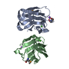
Mass: 40.078 Da / Num. of mol.: 2 / Source method: obtained synthetically / Formula: Ca
#3: Chemical ChemComp-NA / SODIUM ION


Mass: 22.990 Da / Num. of mol.: 2 / Source method: obtained synthetically / Formula: Na
#4: Chemical ChemComp-BTB / 2-[BIS-(2-HYDROXY-ETHYL)-AMINO]-2-HYDROXYMETHYL-PROPANE-1,3-DIOL / BIS-TRIS BUFFER / Bis-tris methane


Mass: 209.240 Da / Num. of mol.: 1 / Source method: obtained synthetically / Formula: C8H19NO5 / Comment: pH buffer*YM
#5: Water ChemComp-HOH / water / Water


Mass: 18.015 Da / Num. of mol.: 498 / Source method: isolated from a natural source / Formula: H2O
Sequence detailsI944V IS A NATURAL VARIANT

-
Experimental details

-
Experiment

ExperimentMethod: X-RAY DIFFRACTION

-
Sample preparation

CrystalDensity Matthews: 1.88 Å3/Da / Density % sol: 34.23 % / Description: NONE

-
Data collection

DiffractionMean temperature: 113.15 K
Diffraction sourceSource: ROTATING ANODE / Wavelength: 1.5418
RadiationProtocol: SINGLE WAVELENGTH / Monochromatic (M) / Laue (L): M / Scattering type: x-ray
Radiation wavelengthWavelength: 1.5418 Å / Relative weight: 1
ReflectionResolution: 1.4→19.74 Å / Num. obs: 109314 / % possible obs: 99.1 % / Observed criterion σ(I): 2 / Redundancy: 7.08 % / Rmerge(I) obs: 0.06 / Net I/σ(I): 15.7
Reflection shellResolution: 1.4→1.45 Å / Redundancy: 4.16 % / Rmerge(I) obs: 0.38 / Mean I/σ(I) obs: 3.2 / % possible all: 91.6

-
Processing

SoftwareName: REFMAC / Version: 5.2.0019 / Classification: refinement
RefinementMethod to determine structure: SIR
Starting model: NONE

Resolution: 1.45→20 Å / Cor.coef. Fo:Fc: 0.97 / Cor.coef. Fo:Fc free: 0.951 / SU B: 3.335 / SU ML: 0.058 / Cross valid method: THROUGHOUT / ESU R: 0.085 / ESU R Free: 0.079 / Stereochemistry target values: MAXIMUM LIKELIHOOD / Details: HYDROGENS HAVE BEEN ADDED IN THE RIDING POSITIONS.
RfactorNum. reflection% reflectionSelection details
Rfree0.226 2651 5.1 %RANDOM
Rwork0.174 ---
obs0.176 49260 99.7 %-
Solvent computationIon probe radii: 0.8 Å / Shrinkage radii: 0.8 Å / VDW probe radii: 1.4 Å / Solvent model: MASK
Displacement parametersBiso mean: 12.19 Å2
Baniso -1Baniso -2Baniso -3
1--0.04 Å20 Å20 Å2
2--0.48 Å20 Å2
3----0.44 Å2
Refinement stepCycle: LAST / Resolution: 1.45→20 Å
ProteinNucleic acidLigandSolventTotal
Num. atoms2192 0 18 498 2708
Refine LS restraints
Refine-IDTypeDev idealDev ideal targetNumber
X-RAY DIFFRACTIONr_bond_refined_d0.0130.0222277
X-RAY DIFFRACTIONr_bond_other_d0.0010.021553
X-RAY DIFFRACTIONr_angle_refined_deg1.461.9523083
X-RAY DIFFRACTIONr_angle_other_deg0.8233801
X-RAY DIFFRACTIONr_dihedral_angle_1_deg6.2895287
X-RAY DIFFRACTIONr_dihedral_angle_2_deg37.88425.728103
X-RAY DIFFRACTIONr_dihedral_angle_3_deg11.00715401
X-RAY DIFFRACTIONr_dihedral_angle_4_deg6.448156
X-RAY DIFFRACTIONr_chiral_restr0.1070.2332
X-RAY DIFFRACTIONr_gen_planes_refined0.0060.022540
X-RAY DIFFRACTIONr_gen_planes_other0.0010.02452
X-RAY DIFFRACTIONr_nbd_refined0.1960.2462
X-RAY DIFFRACTIONr_nbd_other0.2010.21677
X-RAY DIFFRACTIONr_nbtor_refined0.1770.21102
X-RAY DIFFRACTIONr_nbtor_other0.0840.21227
X-RAY DIFFRACTIONr_xyhbond_nbd_refined0.1870.2374
X-RAY DIFFRACTIONr_xyhbond_nbd_other
X-RAY DIFFRACTIONr_metal_ion_refined
X-RAY DIFFRACTIONr_metal_ion_other
X-RAY DIFFRACTIONr_symmetry_vdw_refined0.2050.210
X-RAY DIFFRACTIONr_symmetry_vdw_other0.1930.241
X-RAY DIFFRACTIONr_symmetry_hbond_refined0.1620.259
X-RAY DIFFRACTIONr_symmetry_hbond_other
X-RAY DIFFRACTIONr_symmetry_metal_ion_refined
X-RAY DIFFRACTIONr_symmetry_metal_ion_other
X-RAY DIFFRACTIONr_mcbond_it1.2331.51388
X-RAY DIFFRACTIONr_mcbond_other
X-RAY DIFFRACTIONr_mcangle_it1.79622233
X-RAY DIFFRACTIONr_mcangle_other
X-RAY DIFFRACTIONr_scbond_it2.6853949
X-RAY DIFFRACTIONr_scbond_other
X-RAY DIFFRACTIONr_scangle_it3.5874.5846
X-RAY DIFFRACTIONr_scangle_other
X-RAY DIFFRACTIONr_long_range_B_refined
X-RAY DIFFRACTIONr_long_range_B_other
X-RAY DIFFRACTIONr_rigid_bond_restr
X-RAY DIFFRACTIONr_sphericity_free
X-RAY DIFFRACTIONr_sphericity_bonded
LS refinement shellResolution: 1.45→1.49 Å / Total num. of bins used: 20
RfactorNum. reflection% reflection
Rfree0.46 207 -
Rwork0.358 3565 -
obs--99.71 %

+
About Yorodumi

-
News

-
Feb 9, 2022. New format data for meta-information of EMDB entries

New format data for meta-information of EMDB entries

  • Version 3 of the EMDB header file is now the official format.
  • The previous official version 1.9 will be removed from the archive.

Related info.:EMDB header

External links:wwPDB to switch to version 3 of the EMDB data model

-
Aug 12, 2020. Covid-19 info

Covid-19 info

URL: https://pdbj.org/emnavi/covid19.php

New page: Covid-19 featured information page in EM Navigator.

Related info.:Covid-19 info / Mar 5, 2020. Novel coronavirus structure data

+
Mar 5, 2020. Novel coronavirus structure data

Novel coronavirus structure data

Related info.:Yorodumi Speices / Aug 12, 2020. Covid-19 info

External links:COVID-19 featured content - PDBj / Molecule of the Month (242):Coronavirus Proteases

+
Jan 31, 2019. EMDB accession codes are about to change! (news from PDBe EMDB page)

EMDB accession codes are about to change! (news from PDBe EMDB page)

  • The allocation of 4 digits for EMDB accession codes will soon come to an end. Whilst these codes will remain in use, new EMDB accession codes will include an additional digit and will expand incrementally as the available range of codes is exhausted. The current 4-digit format prefixed with “EMD-” (i.e. EMD-XXXX) will advance to a 5-digit format (i.e. EMD-XXXXX), and so on. It is currently estimated that the 4-digit codes will be depleted around Spring 2019, at which point the 5-digit format will come into force.
  • The EM Navigator/Yorodumi systems omit the EMD- prefix.

Related info.:Q: What is EMD? / ID/Accession-code notation in Yorodumi/EM Navigator

External links:EMDB Accession Codes are Changing Soon! / Contact to PDBj

+
Jul 12, 2017. Major update of PDB

Major update of PDB

  • wwPDB released updated PDB data conforming to the new PDBx/mmCIF dictionary.
  • This is a major update changing the version number from 4 to 5, and with Remediation, in which all the entries are updated.
  • In this update, many items about electron microscopy experimental information are reorganized (e.g. em_software).
  • Now, EM Navigator and Yorodumi are based on the updated data.

External links:wwPDB Remediation / Enriched Model Files Conforming to OneDep Data Standards Now Available in the PDB FTP Archive

-
Yorodumi

Thousand views of thousand structures

  • Yorodumi is a browser for structure data from EMDB, PDB, SASBDB, etc.
  • This page is also the successor to EM Navigator detail page, and also detail information page/front-end page for Omokage search.
  • The word "yorodu" (or yorozu) is an old Japanese word meaning "ten thousand". "mi" (miru) is to see.

Related info.:EMDB / PDB / SASBDB / Comparison of 3 databanks / Yorodumi Search / Aug 31, 2016. New EM Navigator & Yorodumi / Yorodumi Papers / Jmol/JSmol / Function and homology information / Changes in new EM Navigator and Yorodumi

Read more